
The body of a multicellular animal is made up of billions of cells that work together to perform essential life processes. However, individual cells usually do not function independently. Cells with similar structures and specialized functions become organized into groups called tissues. These tissues combine to form organs, while organs work together as organ systems.
Animal tissues are therefore an important level of organization between cells and organs. They provide structural support, protection, movement, communication, transport, and coordination within the animal body.
In animals, tissues are highly specialized because different cells perform different functions. For example, epithelial tissue protects body surfaces, connective tissue provides support and transport, muscular tissue produces movement, and nervous tissue receives and transmits signals.
Understanding the types of animal tissues, their structure, functions, and examples is fundamental to zoology, anatomy, physiology, and histology.
What Are Animal Tissues?
An animal tissue is a group of structurally similar or functionally related cells, along with their associated extracellular material, that work together to perform one or more specific functions.
The cells within a tissue may be similar in appearance and function, although some tissues contain different cell types that cooperate to perform a common function.
For example, blood is classified as a connective tissue even though it contains different components, including red blood cells, white blood cells, platelets, and plasma. Together, these components perform important functions such as transport, defense, and clotting.
How Are Tissues Organized?
The organization of the animal body can be represented as:
Cells → Tissues → Organs → Organ Systems → Organism
For example:
Muscle cells → Muscular tissue → Heart → Circulatory system → Animal
This organization allows cells to become specialized and work together efficiently.
Classification of Animal Tissues
Based on their structure and functions, animal tissues are broadly divided into four major types:
- Epithelial tissue – covers surfaces and lines body cavities and organs.
- Connective tissue – supports, connects, binds, and transports materials.
- Muscular tissue – produces movement through contraction.
- Nervous tissue – receives, processes, and transmits signals.
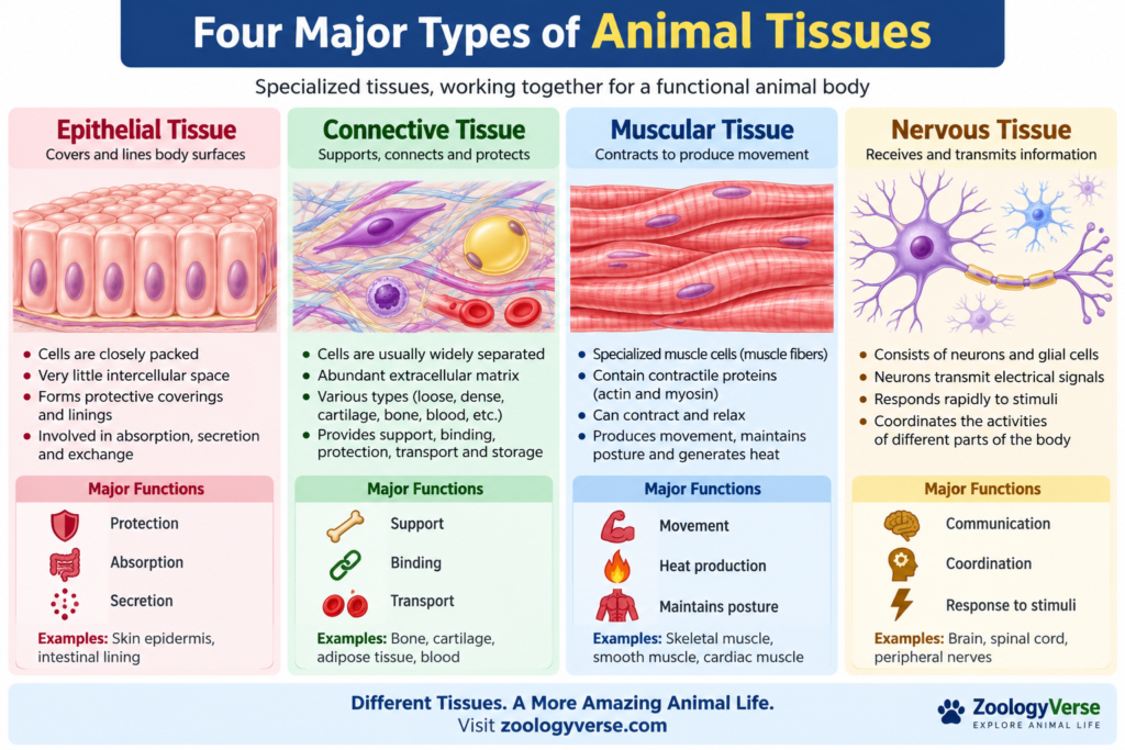
These four major animal tissue types differ considerably in their cellular arrangement, extracellular material, functions, and locations within the body.
We will examine each type in detail in the following sections.
Epithelial Tissue
Epithelial tissue is one of the four major types of animal tissues. It forms the outer covering of the body and lines internal organs, body cavities, blood vessels, and ducts. The cells are closely packed together, leaving very little space between adjacent cells.
The main functions of epithelial tissue include protection, absorption, secretion, filtration, and exchange of substances.
Characteristics of Epithelial Tissue
Important characteristics include:
- Cells are closely packed with very little intercellular space.
- Cells are arranged in one or more continuous layers.
- The tissue rests on a basement membrane.
- Epithelial tissue generally has no blood vessels of its own and receives nutrients by diffusion from underlying tissues.
- Cells are connected by specialized cell junctions.
- The tissue can have a high capacity for repair and regeneration. Many epithelial tissues have a strong capacity for regeneration, which depends on controlled cell division.
- Its structure is closely related to its particular function.
Types of Epithelial Tissue
Based on the number of cell layers, epithelial tissue can be classified into:
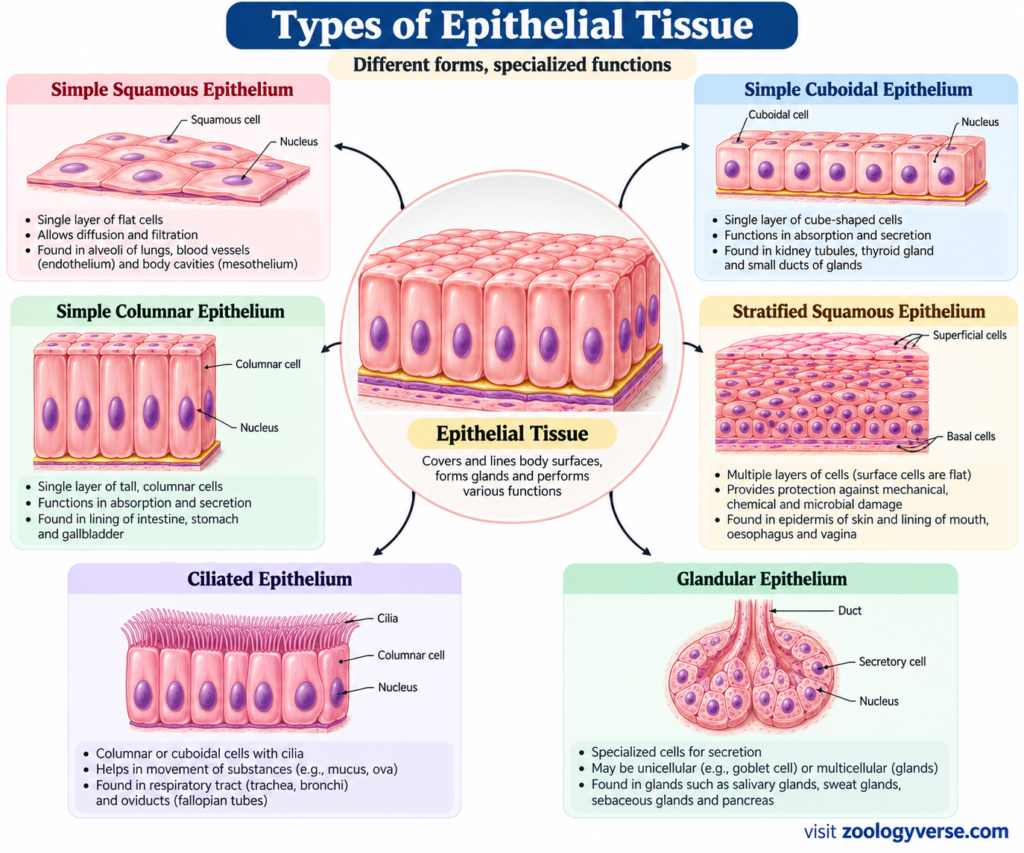
- Simple epithelium – consists of a single layer of cells.
- Stratified epithelium – consists of two or more layers of cells.
Based on the shape of the cells, epithelial cells may be:
- Squamous – thin and flattened
- Cuboidal – approximately cube-shaped
- Columnar – tall and elongated
These characteristics can occur in different combinations, producing several types of epithelial tissue in animals.
1. Simple Squamous Epithelium
Simple squamous epithelium consists of a single layer of thin, flattened cells. Because the cells are very thin, substances can pass across the tissue relatively easily.
Location: Alveoli of lungs, lining of blood vessels, and Bowman’s capsule of the kidney.
Functions: Diffusion, filtration, and reducing friction.
2. Simple Cuboidal Epithelium
This tissue consists of a single layer of cube-shaped cells with centrally located nuclei.
Location: Kidney tubules and ducts of several glands.
Functions: Absorption and secretion.
3. Simple Columnar Epithelium
Simple columnar epithelium consists of a single layer of tall, column-shaped cells. Some cells may contain microvilli or cilia depending on their location and function.
Location: Lining of much of the digestive tract.
Functions: Absorption and secretion.
4. Stratified Squamous Epithelium
Stratified squamous epithelium contains several layers of cells, with flattened cells at the exposed surface.
It occurs in two major forms:
- Keratinized stratified squamous epithelium: Found in the epidermis of the skin and provides protection against abrasion and water loss.
- Non-keratinized stratified squamous epithelium: Found in areas such as the mouth and esophagus where protection is needed but a dry, keratinized surface is not present.
5. Ciliated Epithelium
Ciliated epithelial cells possess tiny hair-like structures called cilia on their free surface. Coordinated movement of cilia helps move materials across the epithelial surface.
Location: Respiratory passages and parts of the female reproductive tract.
Functions: Movement of mucus, particles, or other materials.
6. Glandular Epithelium
Glandular cells contain specialized organelles, including the Golgi body, that participate in processing and secretion.
It forms glands that produce substances such as mucus, enzymes, sweat, and hormones.
Glands may be broadly classified into:
- Exocrine glands – release their secretions through ducts.
- Endocrine glands – release hormones into the surrounding tissue and blood.
Functions of Epithelial Tissue
| Function | Example |
|---|---|
| Protection | Epidermis of skin |
| Absorption | Intestinal epithelium |
| Secretion | Glandular epithelium |
| Diffusion | Alveolar epithelium |
| Filtration | Kidney epithelium |
| Movement of substances | Ciliated epithelium |
| Sensory reception | Specialized sensory epithelium |
Thus, epithelial tissue provides an important interface between the animal’s body and its internal or external environment. Its specialized structure allows it to perform different functions efficiently.
Connective Tissue
Connective tissue is one of the four major types of animal tissues. It connects, supports, protects, and separates different parts of the body. Unlike epithelial tissue, connective tissue generally contains more extracellular material (matrix) between its cells.
The matrix consists of a ground substance and protein fibers, and its composition varies according to the function of the tissue. This variation gives connective tissue its wide range of forms and functions.
Characteristics of Connective Tissue
The important characteristics of connective tissue include:
- Cells are generally widely separated from one another.
- A considerable amount of extracellular matrix is present.
- The matrix may be fluid, gel-like, flexible, or hard.
- The matrix contains different types of protein fibers.
- Most connective tissues have a blood supply, although some, such as cartilage, have limited direct blood supply.
- It provides support, binding, protection, transport, storage, and defense.
Types of Connective Tissue
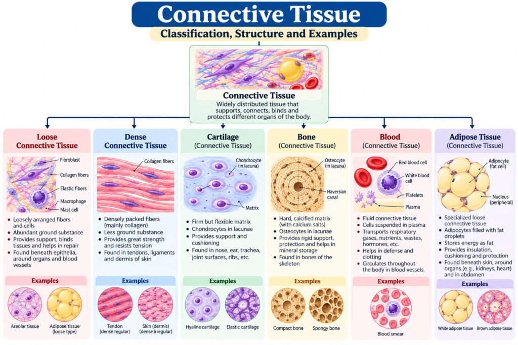
Connective tissue can be broadly divided into:
- Connective tissue proper
- Supporting connective tissue
- Fluid connective tissue
1. Connective Tissue Proper
Connective tissue proper includes loose connective tissue and dense connective tissue.
Loose Connective Tissue
Loose connective tissue contains relatively fewer fibers and more ground substance.
Major types include:
- Areolar tissue
- Adipose tissue
- Reticular tissue
Areolar Tissue
Areolar tissue is a loose connective tissue containing various cells and fibers embedded in a soft matrix.
Location: Beneath the skin, around blood vessels, nerves, and organs.
Functions: Binding tissues, packing spaces, and providing support.
Adipose Tissue
Adipose tissue is composed mainly of adipocytes, or fat cells, which store energy in the form of fat.
Location: Beneath the skin and around organs such as the kidneys.
Functions:
- Energy storage
- Insulation
- Protection and cushioning of organs
Reticular Tissue
Reticular tissue contains a network of reticular fibers that forms a supportive framework for certain organs.
Location: Lymph nodes, spleen, and bone marrow.
Function: Provides structural support for cells within these organs.
Dense Connective Tissue
Dense connective tissue contains a greater proportion of fibers and provides strong mechanical support.
It includes:
- Dense regular connective tissue
- Dense irregular connective tissue
- Elastic connective tissue
Dense Regular Connective Tissue
The collagen fibers are arranged mainly in parallel bundles.
Examples: Tendons and many ligaments.
Function: Provides strong attachment and resistance to tension in a particular direction.
Dense Irregular Connective Tissue
The collagen fibers are arranged in different directions, allowing the tissue to withstand stress from multiple directions.
Location: Dermis of the skin and protective coverings around certain organs.
Function: Provides strength and structural support.
Elastic Connective Tissue
This tissue contains abundant elastic fibers, allowing it to stretch and return toward its original shape.
Location: Walls of certain large arteries and some elastic ligaments.
Function: Provides elasticity and recoil.
2. Supporting Connective Tissue
Supporting connective tissue provides structural support to the body. It mainly includes cartilage and bone.
Cartilage
Cartilage is a firm but flexible connective tissue. Its cells, called chondrocytes, are located within spaces called lacunae in the extracellular matrix.
Major types of cartilage include:
- Hyaline cartilage
- Elastic cartilage
- Fibrocartilage
Functions: Support, flexibility, protection, and smooth movement at joints.
Bone
Bone is a hard connective tissue with a mineralized extracellular matrix. Its principal cells include osteocytes.
Functions of bone include:
- Supporting the body
- Protecting internal organs
- Providing attachment sites for muscles
- Helping movement
- Storing minerals such as calcium and phosphate
- Housing bone marrow involved in blood cell formation
3. Fluid Connective Tissue
Fluid connective tissue has a liquid matrix, allowing substances to be transported throughout the body.
The major examples are:
Blood
Blood consists of plasma and formed elements, including:
- Red blood cells
- White blood cells
- Platelets
Functions: Transport of gases, nutrients, hormones, and waste products; defense; and blood clotting.
Lymph
Lymph is a fluid connective tissue associated with the lymphatic system.
Functions: Helps return tissue fluid to the circulation and participates in immune defense.
Functions of Connective Tissue
The major functions of connective tissue include:
| Function | Example |
|---|---|
| Support | Bone and cartilage |
| Binding | Tendons and ligaments |
| Energy storage | Adipose tissue |
| Transport | Blood |
| Protection | Bone and connective tissue coverings |
| Defense | White blood cells and lymphoid tissues |
| Insulation | Adipose tissue |
| Repair | Various connective tissues |
Key Point
The diversity of connective tissue results largely from differences in its cells, fibers, and extracellular matrix. This is why connective tissue can range from soft areolar tissue to hard bone and fluid blood.
Muscular Tissue
Muscular tissue is one of the four major types of animal tissues. It consists of specialized cells called muscle fibers that can contract and relax. This ability allows muscles to produce movement, maintain posture, stabilize joints, and move substances through internal organs.
The contraction of muscular tissue is mainly associated with the interaction of actin and myosin, two contractile proteins present within muscle cells.
Characteristics of Muscular Tissue
The major characteristics of muscular tissue include:
- It is composed of specialized muscle cells or muscle fibers.
- Muscle cells contain contractile proteins, mainly actin and myosin.
- Contraction occurs when the contractile proteins interact.
- Muscular tissue can respond to appropriate stimuli.
- It helps produce movement of the body and internal organs.
- Depending on the type, contraction may be voluntary or involuntary.
Types of Muscular Tissue
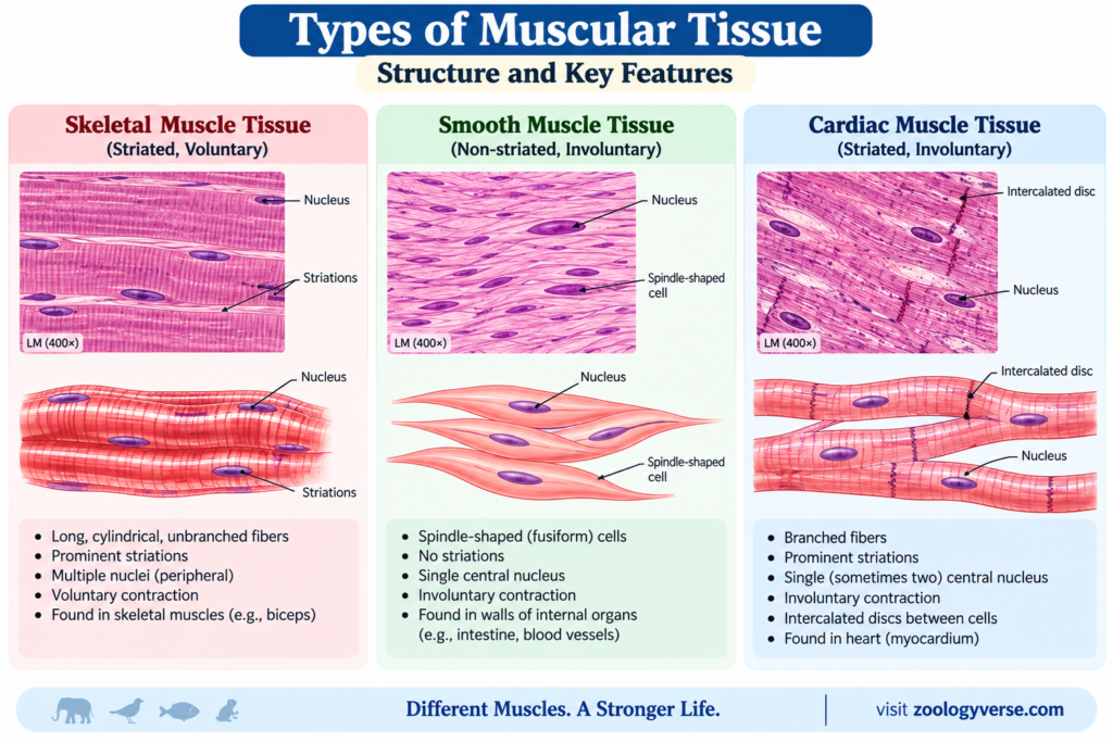
Based on structure, location, and mode of contraction, muscular tissue is divided into three major types:
- Skeletal muscle tissue
- Smooth muscle tissue
- Cardiac muscle tissue
1. Skeletal Muscle Tissue
Skeletal muscle tissue is attached mainly to bones and is responsible for movements of the body and limbs.
The muscle fibers are generally long, cylindrical, and striated, meaning they show alternating light and dark bands when viewed under a microscope. Skeletal muscle fibers usually contain multiple nuclei located near the periphery of the cell.
Muscle cells contain many mitochondria, which provide ATP needed for contraction.
Location: Attached to bones and associated with the skeleton.
Functions:
- Produces voluntary body movements
- Maintains posture
- Stabilizes joints
- Generates heat during contraction
Example: Biceps and triceps muscles.
2. Smooth Muscle Tissue
Smooth muscle tissue is composed of spindle-shaped cells that lack the visible striations characteristic of skeletal and cardiac muscle.
Its contraction is generally involuntary and is controlled largely by the autonomic nervous system, hormones, and local factors.
Location: Walls of organs such as the intestine, stomach, urinary bladder, and blood vessels.
Functions:
- Moves food through the digestive tract
- Regulates the diameter of blood vessels
- Helps empty the urinary bladder
- Produces movements in several internal organs
3. Cardiac Muscle Tissue
Cardiac muscle tissue is found specifically in the wall of the heart. Like skeletal muscle, it is striated, but cardiac muscle cells are generally shorter and branched.
Adjacent cardiac muscle cells are connected by specialized structures called intercalated discs, which help coordinate contraction.
Cardiac muscle contracts involuntarily and continuously throughout life.
Location: Heart wall, particularly the myocardium.
Functions:
- Produces rhythmic contractions of the heart
- Pumps blood through the circulatory system
- Maintains continuous circulation
Comparison of the Three Types of Muscular Tissue
| Feature | Skeletal Muscle | Smooth Muscle | Cardiac Muscle |
|---|---|---|---|
| Striations | Present | Absent | Present |
| Control | Usually voluntary | Involuntary | Involuntary |
| Cell shape | Long and cylindrical | Spindle-shaped | Usually one |
| Nuclei | Usually many | Usually one | Usually one |
| Main location | Attached to bones | Walls of internal organs | Heart |
| Major function | Body movement | Movement of substances within organs | Pumping blood |
Functions of Muscular Tissue
The major functions of muscular tissue include:
- Movement: Skeletal muscles move bones and body parts.
- Posture: Continuous muscle activity helps maintain body position.
- Internal movement: Smooth muscles move substances through organs.
- Blood circulation: Cardiac muscle pumps blood.
- Heat production: Skeletal muscle activity contributes to body heat.
- Regulation: Smooth muscle helps regulate the diameter of blood vessels and other hollow structures.
Key Point
Although all three types of muscular tissue are specialized for contraction, they differ in their structure, location, and control. Skeletal muscle is primarily associated with voluntary movement, whereas smooth and cardiac muscles perform predominantly involuntary functions.
Nervous Tissue
Nervous tissue is one of the four major types of animal tissues and is specialized for receiving, processing, and transmitting information. It forms the basic structural and functional component of the nervous system, including the brain, spinal cord, and peripheral nerves.
Nervous tissue allows animals to detect changes in their internal and external environments and respond appropriately. Its major components are neurons and glial cells (neuroglia).
Characteristics of Nervous Tissue
Important characteristics of nervous tissue include:
- It contains specialized cells called neurons that transmit electrical signals.
- It also contains glial cells, which support and maintain neurons.
- Neurons are highly specialized for communication.
- Many neurons have long cellular processes that allow signals to travel over considerable distances.
- Nervous tissue can respond rapidly to various stimuli.
- It coordinates the activities of different parts of the body.
Components of Nervous Tissue
Nervous tissue mainly consists of two types of cells:
1. Neurons
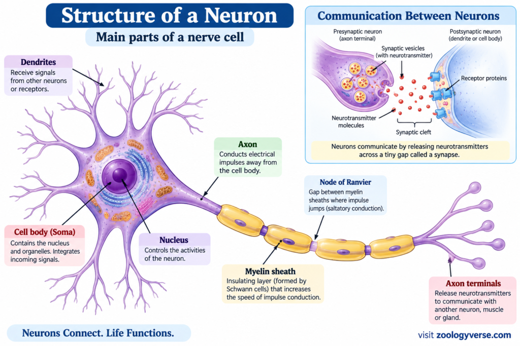
Neurons are the principal signaling cells of nervous tissue. They receive and transmit information using electrical signals and chemical communication.
A typical neuron consists of three main parts:
- Cell body (soma): Contains the nucleus and most of the cell’s organelles.
- Dendrites: Usually receive signals from other cells or sensory receptors.
- Axon: Conducts signals away from the cell body toward other neurons, muscles, or glands.
2. Neuroglia
Neuroglia, or glial cells, are supporting cells of nervous tissue. They do not function primarily as long-distance signaling cells like neurons.
Depending on their type and location, glial cells help:
- Support neurons
- Maintain the chemical environment around neurons
- Provide nutrients and metabolic support
- Protect nervous tissue
- Participate in repair processes
- Produce insulating myelin around many axons
In the central nervous system, important glial cells include astrocytes, oligodendrocytes, microglia, and ependymal cells. In the peripheral nervous system, Schwann cells and satellite cells are important types of glia.
Types of Neurons
Neurons can be classified according to their function.
Sensory Neurons
Sensory neurons carry information from sensory receptors toward the central nervous system.
They are involved in detecting stimuli such as touch, temperature, pressure, and changes in the environment.
Motor Neurons
Motor neurons transmit signals from the central nervous system toward muscles or glands, producing an appropriate response.
Interneurons
Interneurons are mainly located within the central nervous system and connect neurons with one another. They play an important role in processing and integrating information.
Functions of Nervous Tissue
The major functions of nervous tissue include:
| Function | Description |
|---|---|
| Sensory reception | Detects changes in the internal and external environment |
| Signal transmission | Carries information between different parts of the body |
| Integration | Processes and interprets incoming information |
| Coordination | Coordinates activities of organs and organ systems |
| Response | Helps produce appropriate responses to stimuli |
| Regulation | Contributes to control of movement and physiological processes |
Example of Nervous Tissue Function
When a person touches a hot object:
Heat stimulus → Sensory receptors → Sensory neuron → Central nervous system → Motor neuron → Muscle response
This rapid communication helps the body withdraw the hand from the source of heat.
Nervous Tissue in the Body
Nervous tissue is primarily found in:
- Brain
- Spinal cord
- Peripheral nerves
- Sensory structures
Together, these structures form an integrated communication network that enables animals to sense their surroundings, coordinate movement, and regulate many physiological activities.
Key Point
Nervous tissue is specialized for rapid communication and coordination. Neurons transmit information, while glial cells provide essential structural, metabolic, and functional support.
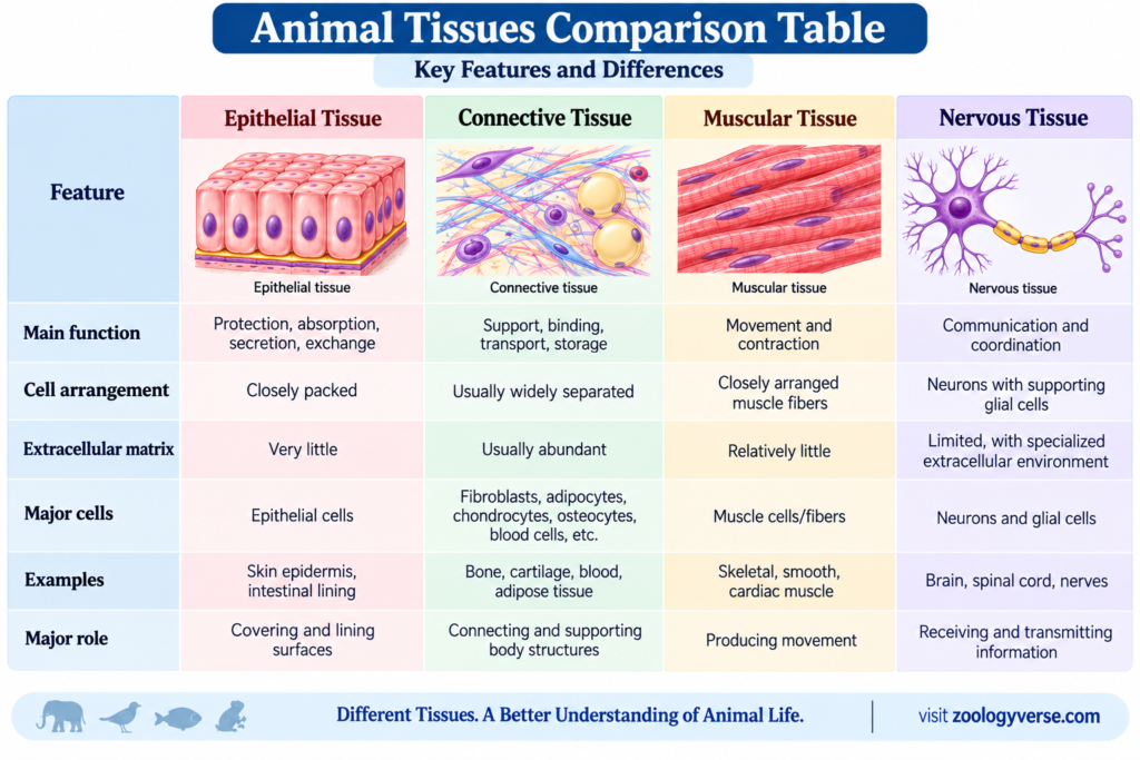
Comparison of the Four Major Types of Animal Tissues
The four major animal tissue types—epithelial, connective, muscular, and nervous tissue—differ in their cellular organization, extracellular matrix, location, and functions. Each type is specialized to perform particular roles, but they work together to maintain the structure and functioning of the animal body.
Functions of Animal Tissues
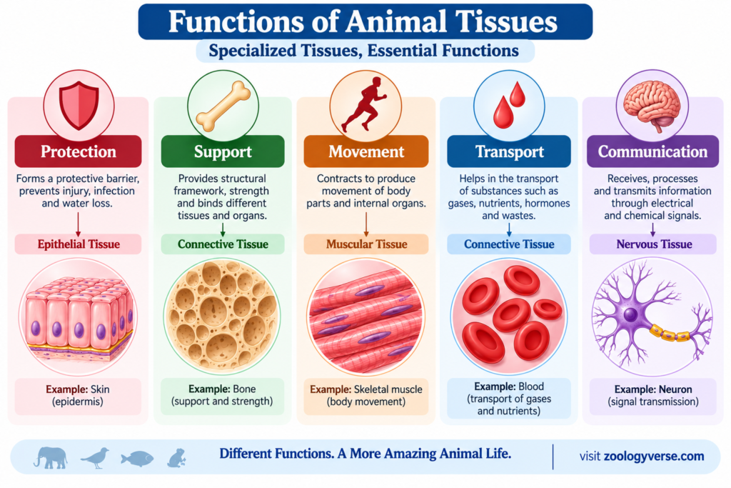
Each tissue type performs specialized functions, but their activities are interconnected.
1. Protection
Epithelial tissue protects the body from:
- Mechanical damage
- Pathogens
- Excessive water loss
- Harmful environmental factors
The epidermis of the skin is an important example of protective epithelial tissue.
2. Support and Connection
Connective tissue provides structural support and connects different parts of the body.
For example:
- Bone supports the body.
- Cartilage provides flexible support.
- Tendons connect muscles to bones.
- Ligaments connect bones to other bones.
3. Movement
Muscular tissue produces movement through contraction.
Skeletal muscles move body parts, while smooth muscles produce movements within internal organs and cardiac muscle drives the pumping action of the heart.
4. Transport
Some connective tissues have important transport functions.
Blood, for example, transports:
- Oxygen
- Carbon dioxide
- Nutrients
- Hormones
- Metabolic waste products
5. Communication and Coordination
Nervous tissue enables rapid communication between different parts of the body.
It receives sensory information, processes signals, and helps coordinate appropriate responses.
6. Secretion and Absorption
Specialized epithelial tissues perform important exchange and secretory functions.
For example:
- Intestinal epithelium participates in nutrient absorption.
- Glandular epithelium produces secretions.
- Respiratory epithelium participates in exchange and protection.
Why Are Animal Tissues Important?
The specialization of tissues allows multicellular animals to perform complex functions efficiently. Instead of every cell carrying out all functions independently, groups of specialized cells perform particular tasks.
For example:
Nervous tissue detects and communicates information →
Muscular tissue produces movement →
Connective tissue provides support →
Epithelial tissue protects and regulates exchange.
Together, these animal tissues contribute to the organization and functioning of organs and organ systems.
Animal Tissues vs Plant Tissues Although both plants and animals are multicellular organisms, their tissues differ because of their different lifestyles and functional requirements.
| Feature | Animal Tissues | Plant Tissues |
|---|---|---|
| Major tissue groups | Epithelial, connective, muscular, nervous | Meristematic and permanent tissues |
| Movement | Muscular tissue enables active movement | No muscular tissue |
| Coordination | Nervous tissue | Mainly chemical and electrical signaling through plant systems |
| Support | Bone, cartilage, connective tissues | Cell walls, vascular and supporting tissues |
| Growth pattern | Generally more limited after maturity in many tissues | Meristematic tissues permit growth throughout life in many regions |
Key Point
The four major types of animal tissues are structurally and functionally specialized. Their coordinated activities form the foundation of organs and organ systems, allowing animals to carry out complex life processes.
Key Takeaways
- Animal tissues are groups of specialized cells that work together to perform specific functions.
- The four major types are epithelial, connective, muscular, and nervous tissue.
- Epithelial tissue covers body surfaces, lines organs and cavities, and performs functions such as protection, absorption, and secretion.
- Connective tissue supports, connects, protects, stores, and transports materials throughout the body.
- Muscular tissue is specialized for contraction and produces body and internal organ movements.
- Nervous tissue receives, processes, and transmits information through neurons and supporting glial cells.
- The structure of each tissue is closely related to its particular function.
- Different tissues combine to form organs, and organs work together as organ systems.
- Understanding animal tissue types, structure, and functions is important in zoology, anatomy, physiology, and histology.
Conclusion
Animal tissues form the basic structural and functional organization of the bodies of multicellular animals. The four major types of animal tissues—epithelial, connective, muscular, and nervous tissue— have distinct structures that allow them to perform specialized functions.
Epithelial tissue provides protection and carries out absorption and secretion, while connective tissue supports, connects, protects, and transports materials. Muscular tissue produces movement through contraction, and nervous tissue enables rapid communication and coordination.
The coordinated activity of these animal tissue types allows cells, organs, and organ systems to function together efficiently. Therefore, understanding their structure, functions, and examples is essential for studying zoology, anatomy, physiology, and histology.
In short:
Cells → Tissues → Organs → Organ Systems → Organism
This organization allows animals to perform increasingly complex biological functions.
References
- Alberts, B. et al. Molecular Biology of the Cell. Garland Science.
- Campbell, N. A. et al. Campbell Biology. Pearson.
- Hickman, C. P. et al. Integrated Principles of Zoology. McGraw-Hill.
- Ross, M. H. & Pawlina, W. Histology: A Text and Atlas. Wolters Kluwer.
- Junqueira, L. C. & Carneiro, J. Junqueira’s Basic Histology: Text and Atlas. McGraw-Hill.
- OpenStax. Anatomy and Physiology 2e — chapters on tissues and body organization.
- Encyclopaedia Britannica. Tissue: Biology.
Recommended Study Resource
JUNQUIRA’S BASIC HISTOLOGY TEXT & ATLAS 17E (IE)
by Anthony L. Mescher (Author)
Integrated Principles of Zoology
by Jr. Hickman, Cleveland P. (Author), Larry S. Roberts (Author), Susan L. Keen (Author), David L. Eisenhour (Author)
As an Amazon Associate I earn from qualifying purchases.
Discover more from Zoologyverse
Subscribe to get the latest posts sent to your email.