
WHAT IS OXIDATIVE PHOSPHORYLATION
Every cell needs a steady supply of usable energy, and for most living things, that energy comes packaged as ATP. Oxidative phosphorylation is the final stage of this energy-harvesting process — the point where the electrons stripped from food molecules during earlier metabolic steps get funneled through a chain of proteins, ultimately powering the production of ATP.
The Chemiosmotic Theory: The Core Idea Behind ATP Production
In simple terms, three things happen in sequence:
- Electrons move along a series of membrane-bound protein complexes toward oxygen, their final destination.
- That electron movement releases energy, which cells use to pump protons across a membrane.
- Protons flow back across that membrane through a specialized enzyme, and that flow drives ATP production.
This entire framework is known as the chemiosmotic theory, first proposed by biochemist Peter Mitchell in 1961.
It remains one of the most important unifying ideas in modern biology, explaining not just mitochondrial energy production but related processes like photosynthesis and even bacterial movement.
In eukaryotic cells, this all takes place inside mitochondria — specifically across their inner membrane, where a series of massive protein machines carry out the work.
Before Mitchell’s theory, scientists struggled to explain exactly how the energy from electron transfer got converted into ATP.
His answer: the cell doesn’t convert that energy directly. Instead, it stores it temporarily as a proton gradient — a difference in proton concentration across the mitochondrial membrane.
Think of it like a dam. Electron transport does the work of pumping protons “uphill” into the space between the mitochondrial membranes, building up potential energy.
When those protons are finally allowed to flow back “downhill,” that flow gets captured by a molecular turbine — ATP synthase — which converts it into chemical energy in the form of ATP.
This coupling between electron flow and ATP production is why the two processes can’t be separated: one cannot happen without the other.
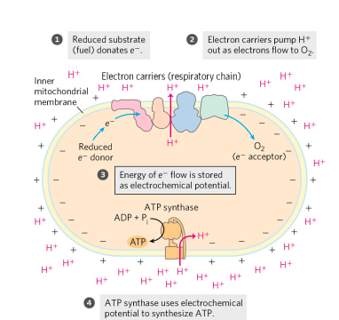
(1) Electrons flow from electron donors (oxidizable substrates) through a chain of membrane-bound carriers to a final electron acceptor with a large reduction potential (molecular oxygen, O2 ).
(2) The free energy made available by this “downhill” (exergonic) electron flow is coupled to the “uphill” transport of protons across a proton-impermeable membrane, conserving the free energy of fuel oxidation as a transmembrane electrochemical potential.
(3) The transmembrane flow of protons back down their concentration gradient through specific protein channels provides the free energy for synthesis of ATP, catalyzed by a membrane protein complex (ATP synthase) that couples proton flow to phosphorylation of ADP.
The Mitochondrial Respiratory Chain
(A) Complex I: NADH to Ubiquinone
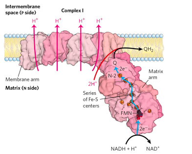
In mammals, Complex I, also called NADH:ubiquinone oxidoreductase or NADH dehydrogenase, is a large enzyme composed of 45 different polypeptide chains, including an FMNcontaining flavoprotein and at least 8 iron-sulfur centers.
Complex I is L shaped, with one arm embedded in the inner membrane and the other extending into the matrix.
Complex I catalyzes two simultaneous and obligately coupled processes: (1) the exergonic transfer to ubiquinone of a hydride ion from NADH and a proton from the matrix, expressed by

and (2) the endergonic transfer of four protons from the matrix to the intermembrane space. Protons are moved against a transmembrane proton gradient in this process.
Complex I is therefore a proton pump driven by the energy of electron transfer.
IRON SULPHUR CENTRE
In iron-sulfur proteins, the iron is present not in heme but in association with inorganic sulfur atoms or with the sulfur atoms of Cys residues in the protein, or both.
These iron-sulfur (Fe-S) centers range from simple structures with a single Fe atom coordinated to four Cys —SH groups to more complex Fe-S centers with two or four Fe atoms.
Rieske iron-sulfur proteins (named after their discoverer, John S. Rieske) are a variation on this theme, in which one Fe atom is coordinated to two His residues rather than two Cys residues.
All iron-sulfur proteins participate in one-electron transfers in which one iron atom of the Fe-S cluster is oxidized or reduced.
At least eight Fe-S proteins function in mitochondrial electron transfer. The reduction potential of Fe-S proteins varies from −0.65 V to +0.45 V, depending on the microenvironment of the iron within the protein.
(B) Complex II: Succinate to Ubiquinone
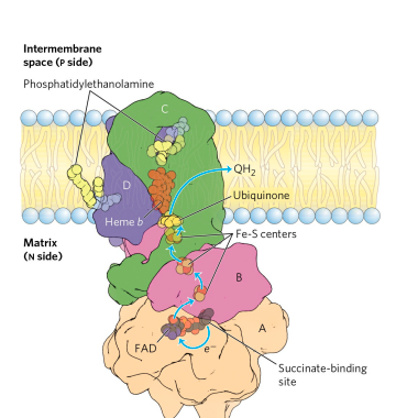
Complex II couples the oxidation of succinate at one site with the reduction of ubiquinone at another site about 40 Å away.
Although smaller and simpler than Complex I, Complex II contains five prosthetic groups of two types and four different protein subunits.
Subunits C and D are integral membrane proteins, each with three transmembrane helices.
They contain a heme group, heme b, and a binding site for Q, the final electron acceptor in the reaction catalyzed by Complex II.
Subunits A and B extend into the matrix; they contain three 2Fe-2S centers, bound FAD, and a binding site for the substrate, succinate.
Although the overall path of electron transfer is long (from the succinate-binding site to FAD, then through the Fe-S centers to the Q-binding site), none of the individual electron-transfer distances exceeds about 11 Å—a reasonable distance for rapid electron transfer.
Electron transfer through Complex II is not accompanied by proton pumping across the inner membrane, although the QH2 produced by succinate oxidation will be used by Complex III to drive proton transfer.
Because Complex II functions in the citric acid cycle, factors that affect its activity (such as the availability of oxidized Q) probably serve to coordinate that cycle with mitochondrial electron transfer.
(C) Complex III: Ubiquinone to Cytochrome c
Electrons from reduced ubiquinone (ubiquinol, QH2 ) pass through two more large protein complexes in the inner mitochondrial membrane before reaching the ultimate electron acceptor, O2 .
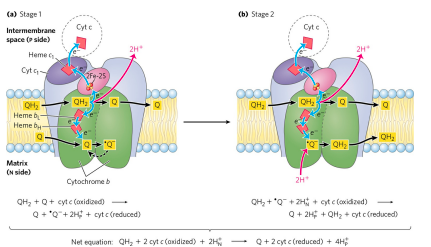
Complex III (also called cytochrome bc1 complex or ubiquinone:cytochrome c oxidoreductase) couples the transfer of electrons from ubiquinol to cytochrome c with the vectorial transport of protons from the matrix to the intermembrane space.
The functional unit of Complex III is a dimer.
Each monomer consists of three proteins central to the action of the complex: cytochrome b, cytochrome c1 , and the Rieske ironsulfur protein. (Several other proteins associated with Complex III in vertebrates are not conserved across the phyla and presumably play subsidiary roles.)
The two cytochrome b monomers surround a cavern in the middle of the membrane, in which ubiquinone is free to move from the matrix side of the membrane (site QN on one monomer) to the intermembrane space (site QP on the other monomer) as it shuttles electrons and protons across the inner mitochondrial membrane.
To account for the role of Q in energy conservation, Mitchell proposed the “Q cycle”. As electrons move from QH2 through Complex III, QH2 is oxidized with the release of protons on one side of the membrane (at QP ), while at the other site (QN), Q is reduced and protons are taken up.
The product of one catalytic site thus becomes the substrate at the second site, and vice versa.
The Q cycle accommodates the switch between the two-electron carrier ubiquinol (the reduced form of ubiquinone) and the one-electron carriers— hemes bL and bH of cytochrome b, and cytochromes c1 and c—and results in the uptake of two protons on the N side and the release of four protons on the P side, per pair of electrons passing through Complex III to cytochrome c.
Two of the protons released on the P side are electrogenic; the other two are electroneutral, balanced by the two charges (electrons) passed to cytochrome c on the P side.
Although the path of electrons through this segment of the respiratory chain is complicated, the net effect of the transfer is simple: QH2 is oxidized to Q, two molecules of cytochrome c are reduced, and two protons are moved from the P side to the N side of the inner mitochondrial membrane.
Cytochrome c is a soluble protein of the intermembrane space, which associates reversibly with the P side of the inner membrane. After its single heme accepts an electron from Complex III, cytochrome c moves in the intermembrane space to Complex IV to donate the electron to a binuclear copper center.
(D) Complex IV: Cytochrome c to O2
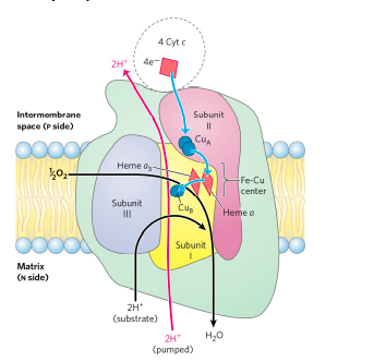
In the final step of the respiratory chain, Complex IV, also called cytochrome oxidase, carries electrons from cytochrome c to molecular oxygen, reducing it to H2O.
Complex IV is a large, dimeric enzyme of the inner mitochondrial membrane,
Subunit II of Complex IV contains two Cu ions complexed with the —SH groups of two Cys residues in a binuclear center that resembles the 2Fe-2S centers of iron-sulfur proteins.
Subunit I contains two heme groups, designated a and a3 , and another copper ion (CuB). Heme a3 and CuB form a second binuclear center that accepts electrons from heme a and transfers them to O2 bound to heme a3 .
Electron transfer through Complex IV is from cytochrome c to the CuA center, to heme a, to the heme a3–CuB center, and finally to O2. For every two electrons passing through this complex, the enzyme consumes two “substrate” H+ from the matrix (N side) in converting ½O2 to H2O. It also uses the energy of this redox reaction to pump two protons outward into the intermembrane space (P side) for each pair of electrons that pass through, adding to the electrochemical potential produced by redox-driven proton transport through Complexes I and III.
This two-electron reduction of ½O2 requires the oxidation of QH2 , which in turn requires oxidation of NADH or succinate.
At Complex IV, O2 is reduced at redox centers that carry only one electron at a time. Normally the incompletely reduced oxygen intermediates remain tightly bound to the complex until completely converted to water, but a small fraction of oxygen intermediates escape.
ATP SYNTHESIS IN OXIDATIVE PHOSPHORYLATION
How is a concentration gradient of protons transformed into ATP? Electron transfer releases, and the proton-motive force conserves, more than enough free energy (about 190 kJ) per “mole” of electron pairs to drive the formation of a mole of ATP, which requires about 50 kJ.
Mitochondrial oxidative phosphorylation therefore poses no thermodynamic problem. But what is the chemical mechanism that couples proton flux with phosphorylation?
THE CHEMIOSMOTIC MODEL
According to the model , the electrochemical energy inherent in the difference in proton concentration and the separation of charge across the inner mitochondrial membrane—the proton-motive force —drives the synthesis of ATP as protons flow passively back into the matrix through a proton pore in ATP synthase.
The overall process is sometimes referred to as “chemiosmotic coupling.” Here, “coupling” refers to the obligate connection between mitochondrial ATP synthesis and electron flow through the respiratory chain; neither of the two processes can proceed without the other.
How ATP Synthase Works
This large enzyme complex of the inner mitochondrial membrane catalyzes the formation of ATP from ADP and Pi , driven by the flow of protons from the P to the N side of the membrane.
ATP synthase, also called Complex V, has two distinct components: F1 , a peripheral membrane protein, and Fo (o denoting oligomycin-sensitive), which is integral to the membrane. F1.
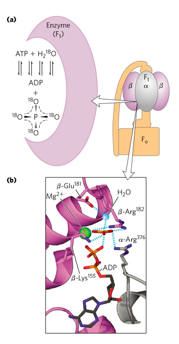
Mitochondrial F1 has nine subunits of five different types, with the composition α3β3γδε. Each of the three β subunits has one catalytic site for ATP synthesis.
The crystallographic determination of the F1. The knoblike portion of F1 is a flattened sphere, 8 nm by 10 nm, consisting of alternating α and β subunits.
The polypeptides that make up the stalk in the F1 crystal structure are asymmetrically arranged, with one domain of the single γ subunit making up a central shaft that passes through F1.
The Fo complex, with its proton pore, is composed of three subunits, a, b, and c, in the proportion ab2cn.
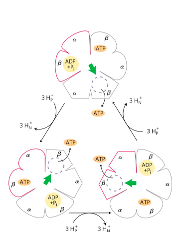
Paul Boyer proposed a rotational catalysis mechanism in which the three active sites of F1 take turns catalyzing ATP synthesis.
A given β subunit starts in the β-ADP conformation, which binds ADP and Pi from the surrounding medium.
The subunit now changes conformation, assuming the β-ATP form that tightly binds and stabilizes ATP, bringing about the ready equilibration of ADP + Pi with ATP on the enzyme surface.
Finally, the subunit changes to the β-empty conformation, which has very low affinity for ATP, and the newly synthesized ATP leaves the enzyme surface.
Another round of catalysis begins when this subunit again assumes the β-ADP form and binds ADP and Pi.
The F1 complex has three nonequivalent adenine nucleotide–binding sites, one for each pair of α and β subunits.
At any given moment, one of these sites is in the β-ATP conformation (which binds ATP tightly), a second is in the β-ADP (loosebinding) conformation, and a third is in the β-empty (very-loose-binding) conformation.
In this view from the N side, the proton-motive force causes rotation of the central shaft—the γ subunit, which comes into contact with each αβ subunit pair in succession.
This produces a cooperative conformational change in which the β-ATP site is converted to the β-empty conformation, and ATP dissociates; the β-ADP site is converted to the β-ATP conformation, which promotes condensation of bound ADP + Pi to form ATP; and the β-empty site becomes a β-ADP site, which loosely binds ADP + Pi entering from the solvent.
This model, based on experimental findings, requires that at least two of the three catalytic sites alternate in activity; ATP cannot be released from one site unless and until ADP and Pi are bound at the other.
Note that the direction of rotation reverses when the ATP synthase is acting as an ATPase One strong prediction of this binding-change model is that the γ subunit should rotate in one direction when FoF1 is synthesizing ATP and in the opposite direction when the enzyme is hydrolyzing ATP.
CONCLUSION
- Chemiosmotic theory provides the intellectual framework for understanding many biological energy transductions, including oxidative phosphorylation and photophosphorylation.
- The energy of electron flow is conserved by the concomitant pumping of protons across the membrane, producing an electrochemical gradient, the proton-motive force.
- In mitochondria, hydride ions removed from substrates (such as αketoglutarate and malate) by NAD-linked dehydrogenases donate electrons to the respiratory chain, which transfers the electrons to molecular O2 , reducing it to H2O.
- Reducing equivalents from NADH are passed through a series of Fe-S centers to ubiquinone, which transfers the electrons to cytochrome b, the first carrier in Complex III.
- In this complex, electrons take two separate paths through two b-type cytochromes and cytochrome c1 to an Fe-S center.
- The Fe-S center passes electrons, one at a time, through cytochrome c and into Complex IV, cytochrome oxidase.
- This copper-containing enzyme, which also contains cytochromes a and a3 , accumulates electrons, then passes them to O2 , reducing it to H2O.
- Some electrons enter this chain of carriers through alternative paths.
- Succinate is oxidized by succinate dehydrogenase (Complex II), which contains a flavoprotein that passes electrons through several Fe-S centers to ubiquinone.
- Electrons derived from the oxidation of fatty acids pass to ubiquinone via the electron-transferring flavoprotein.
- The oxidation of glycerol phosphate and of dihydroorotate also sends electrons into the respiratory chain at the level of QH2 .
- Potentially harmful reactive oxygen species produced in mitochondria are inactivated by a set of protective enzymes, including superoxide dismutase and glutathione peroxidase.
- Low levels of ROS serve as signals coordinating mitochondrial oxidative phosphorylation with other metabolic pathways.
- Plants, fungi, and unicellular eukaryotes have, in addition to the typical path for electron transfer coupled to ATP synthesis, an alternative, uncoupled pathway that recycles excess NADH to NAD+ .
- The flow of electrons through Complexes I, III, and IV results in pumping of protons across the inner mitochondrial membrane, making the matrix alkaline relative to the intermembrane space. This proton gradient provides the energy, in the form of the proton-motive force, for ATP synthesis from ADP and Pi by ATP synthase (FoF1 complex) in the inner membrane.
- ATP synthase carries out “rotational catalysis,” in which the flow of protons through Fo causes each of three nucleotide-binding sites in F1 to cycle from (ADP + Pi )–bound to ATP-bound to empty conformations.
- ATP formation on the enzyme requires little energy; the role of the protonmotive force is to push ATP from its binding site on the synthase.
- The ratio of ATP synthesized per ½O2 reduced to H2O (the P/O ratio) is about 2.5 when electrons enter the respiratory chain at Complex I, and 1.5 when electrons enter at ubiquinone. This ratio varies among species, depending on the number of c subunits in the Fo complex.
- Energy conserved in a proton gradient can drive solute transport uphill across a membrane.
Frequently Asked Questions
What is oxidative phosphorylation in simple terms?
It’s the process cells use to make most of their ATP. Electrons pulled from food molecules travel through a chain of proteins in the mitochondria, and the energy released along the way is used to build ATP.
Where does oxidative phosphorylation take place?
In eukaryotic cells, it happens across the inner mitochondrial membrane. The electron transport chain complexes and ATP synthase are all embedded there.
What is the difference between oxidative phosphorylation and substrate-level phosphorylation?
Substrate-level phosphorylation makes ATP directly during a single enzymatic reaction, without needing oxygen or a membrane gradient. Oxidative phosphorylation is indirect — it relies on an electron transport chain and a proton gradient, and it requires oxygen as the final electron acceptor.
What is the difference between oxidative phosphorylation and photophosphorylation?
Both use the same chemiosmotic principle — a proton gradient driving ATP synthase. Oxidative phosphorylation gets its electrons from breaking down food molecules and happens in mitochondria. Photophosphorylation gets its electrons from light-excited molecules and happens in chloroplasts during photosynthesis.
How much ATP does oxidative phosphorylation produce?
Estimates vary by source and by which shuttle system a cell uses, but oxidative phosphorylation typically accounts for roughly 26–28 of the ~30–32 ATP molecules generated from a single glucose molecule — far more than glycolysis produces on its own.
What happens if oxidative phosphorylation is blocked?
Cells lose their main source of ATP. Certain toxins and drugs work exactly this way — some block specific complexes in the electron transport chain, while others (called uncouplers) let protons leak across the membrane without passing through ATP synthase, wasting the gradient as heat instead of ATP.
Why does oxidative phosphorylation need oxygen?
Oxygen acts as the final electron acceptor at Complex IV. Without it, electrons have nowhere to go, the whole chain backs up, and proton pumping — and therefore ATP synthesis — stops.
External Links (Authoritative Sources)
- Biochemistry, Electron Transport Chain — StatPearls, NCBI Bookshelf
- Biochemistry, Oxidative Phosphorylation — StatPearls, NCBI Bookshelf
- The Mechanism of Oxidative Phosphorylation — The Cell: A Molecular Approach, NCBI Bookshelf
- Oxidative Phosphorylation: Regulation and Role in Cellular and Systemic Metabolism — PMC
See Other Posts Also
- GLYCOLYSIS
- STORAGE LIPIDS (TRIACYLGYCEROL)
- MEMBRANE LIPIDS
- Citric Acid Cycle: 8 Steps, Diagram & Energy Yield Explained
Discover more from Zoologyverse
Subscribe to get the latest posts sent to your email.