
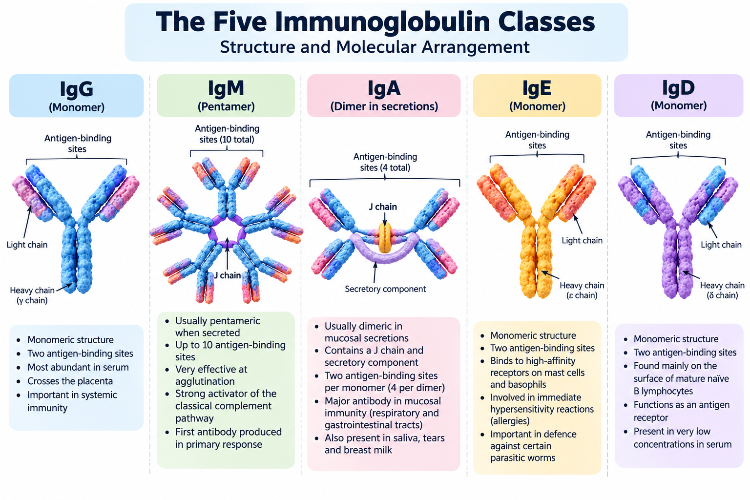
The five types of immunoglobulins found in humans are IgG, IgM, IgA, IgE and IgD. Although they share a common basic molecular design, they differ in their heavy-chain structure, distribution in the body, ability to activate immune mechanisms and specific biological functions.
Immunoglobulins, commonly known as antibodies, are specialized glycoproteins produced by plasma cells, which develop from activated B lymphocytes. They play a central role in adaptive immunity by recognizing specific antigens and helping the body defend itself against pathogens, toxins and other foreign substances.
Each immunoglobulin class has a distinct role in immune protection. IgG is the predominant antibody in serum and contributes to neutralization, opsonization and long-term protection. IgM is commonly the first antibody class secreted during a primary immune response and is particularly effective at agglutination and complement activation. IgA protects mucosal surfaces, including the respiratory and gastrointestinal tracts, while IgE participates in immediate allergic reactions and defense against certain parasitic worms. IgD primarily functions as a membrane-bound antigen receptor on mature naive B cells.
Understanding the different antibody classes helps explain how the immune system provides protection in blood, tissues and mucosal secretions. These differences are also important in clinical diagnosis, immunological research, vaccine studies and the development of antibody-based therapies.
In this article, we will examine the structure, distribution, major functions and biological significance of each immunoglobulin class, followed by a comparison table and exam-oriented key points.
What Are Immunoglobulins?
Immunoglobulins (Igs) are antigen-specific glycoproteins produced in a secreted form by plasma cells and expressed in a membrane-bound form by B lymphocytes. They are essential components of humoral immunity, the branch of adaptive immunity that uses antibodies to recognize antigens and help control infections.
Immunoglobulins are produced after B cells develop the ability to recognize particular antigens. Following activation, some B cells differentiate into plasma cells that secrete large quantities of antibodies, while others become memory B cells that support faster responses during subsequent exposure to the same antigen.
Immunoglobulins are specialised proteins that recognise antigens and contribute to antibody-mediated immunity. To understand their molecular organisation in detail, read our guide to antibody structure.
Basic Characteristics of Immunoglobulins
The major characteristics of immunoglobulins include:
- Antigen specificity: Each antibody has binding sites that recognize particular molecular features, called epitopes, on an antigen.
- Glycoprotein nature: Immunoglobulins consist of amino acid chains with attached carbohydrate groups that can influence their stability and biological activity.
- Specialized molecular structure: The five major classes share a common basic antibody design but differ in their heavy-chain constant regions and structural organization.
- Humoral immune defense: Antibodies can neutralize toxins and pathogens, promote phagocytosis and recruit other immune mechanisms.
- Different distributions: Individual classes occur in different proportions in blood, tissues, mucosal secretions and other biological environments.
- B-cell receptor activity: Membrane-bound immunoglobulins recognize antigens and participate in initiating B-cell activation.
How Are Immunoglobulins Produced?
Immunoglobulin production begins with B lymphocytes, which develop in the bone marrow in humans. During development, gene rearrangements generate diverse antigen-binding specificities, allowing different B cells to recognize different antigens.
When an appropriate antigen activates a B cell, that cell may undergo clonal expansion. Some of its descendants differentiate into antibody-secreting plasma cells, while others become memory B cells.
In many responses to protein antigens, helper T cells provide signals that support B-cell proliferation, antibody production, affinity maturation and class switching. Class switching allows an activated B cell to produce a different immunoglobulin class while generally retaining the antigen specificity of its original variable region.
Immunoglobulins and Humoral Immunity
Humoral immunity protects the body through antibodies present in blood, lymph and other extracellular fluids. After binding to their targets, antibodies can prevent pathogens from attaching to host cells, neutralize harmful toxins, promote the uptake of microorganisms by phagocytes or activate complement proteins.
Different immunoglobulin classes are adapted to different environments and tasks. For example, IgA is especially important at mucosal surfaces, while IgG provides extensive protection in blood and tissues. IgE participates in mast-cell-mediated responses, and membrane-bound IgM and IgD help naive B cells recognize antigens.
For a detailed explanation of the molecular design shared by antibodies, see our related article, Antibody Structure: Parts, Regions and Functions.
Classification of Immunoglobulins
Immunoglobulins are classified into five major classes: IgG, IgM, IgA, IgE and IgD. This classification is based primarily on differences in the constant region of their heavy chains. Although all five classes recognize specific antigens, they differ in their molecular structure, distribution in the body and biological functions.
Immunoglobulins are important components of adaptive immunity, which works alongside innate immune defences to protect the body. Learn more in our article on types of immunity.
Basis of Classification
The heavy chain of an antibody determines its immunoglobulin class. Each class has a characteristic type of heavy chain, represented by a Greek letter.
| Immunoglobulin class | Heavy-chain type | General structural form |
|---|---|---|
| IgG | Gamma (γ) | Monomer |
| IgM | Mu (μ) | Usually a pentamer when secreted |
| IgA | Alpha (α) | Monomer in serum; often a dimer in secretions |
| IgE | Epsilon (ε) | Monomer |
| IgD | Delta (δ) | Monomer |
These structural forms describe the typical secreted antibodies. Membrane-bound immunoglobulins on B lymphocytes can have different arrangements; for example, membrane-bound IgM is monomeric and functions as part of the B-cell receptor.
Structural Differences Among Immunoglobulin Classes
The five immunoglobulin classes share the basic antibody structure but differ in their heavy-chain constant regions and the way their molecules are assembled.
- IgG: A monomer consisting of two heavy chains and two light chains. Humans have four subclasses: IgG1, IgG2, IgG3 and IgG4.
- IgM: Secreted IgM is usually composed of five antibody units joined together. This arrangement provides multiple antigen-binding sites and makes IgM particularly effective at agglutination and complement activation.
- IgA: Exists mainly as a monomer in blood serum. In many mucosal secretions, including saliva, tears and intestinal secretions, it commonly occurs as a dimer called secretory IgA. Human IgA has two subclasses: IgA1 and IgA2.
- IgE: A monomeric antibody with a role in allergic reactions and defence against certain parasitic worms.
- IgD: A monomeric antibody found mainly on the surface of mature naïve B lymphocytes, where it participates in antigen recognition.
Distribution and Functional Specialisation
Each class is adapted to particular immune functions and locations.
| Class | Major location or distribution | Principal role |
|---|---|---|
| IgG | Blood, extracellular fluid and tissues | Neutralisation, opsonisation and placental transfer |
| IgM | Blood and lymph; also on B-cell surfaces | Early antibody response, agglutination and complement activation |
| IgA | Mucosal secretions and blood | Protection of mucosal surfaces |
| IgE | Bound to mast cells and basophils; present at low concentrations in serum | Allergic reactions and defence against helminths |
| IgD | Mainly on naïve B-cell surfaces | B-cell antigen recognition and activation |
These roles are not completely exclusive. For example, several immunoglobulin classes can neutralise pathogens, but they differ in where they act and how effectively they carry out particular immune functions.
Why Are There Five Immunoglobulin Classes?
The existence of five immunoglobulin classes enables the immune system to respond to different types of threats in different body compartments. IgM is particularly important in early responses, IgG provides extensive protection in blood and tissues, and IgA protects mucosal surfaces. IgE participates in responses against certain parasites and in immediate allergic reactions, while IgD is primarily associated with B-cell function.
The production of different classes is regulated by B-cell development and activation. During an immune response, activated B cells may undergo class-switch recombination, changing the heavy-chain class of the antibody they produce while retaining the same antigen specificity, provided the variable region remains unchanged.
The detailed functions of each class are discussed in the following sections, beginning with IgG.
IgG: Structure, Distribution and Functions
Immunoglobulin G (IgG) is the most abundant immunoglobulin in human blood serum and plays a major role in long-term protection against pathogens. It is particularly important in secondary immune responses, when the immune system encounters an antigen it has previously recognised. IgG helps neutralise toxins and viruses, promotes the destruction of microorganisms and provides passive immunity to the developing foetus through the placenta.
Structure of IgG
IgG is a monomeric immunoglobulin, meaning that it consists of a single antibody unit. Like other conventional antibody molecules, it contains two identical heavy chains and two identical light chains linked by disulfide bonds.
The heavy chains of IgG are called gamma (γ) chains. Each molecule has two antigen-binding sites, allowing it to bind two identical epitopes when their arrangement permits.
Important structural features of IgG include:
- Two antigen-binding sites: Recognise specific epitopes on antigens.
- Fab regions: Contain the antigen-binding portions of the molecule.
- Fc region: Interacts with Fc receptors on immune cells and helps recruit effector mechanisms.
- Hinge region: Provides flexibility for antigen binding.
- Disulfide bonds: Help stabilise the association between the heavy and light chains and between the heavy chains.
For a detailed explanation of these structural regions, see the article on Antibody Structure.
Distribution of IgG
IgG is found mainly in blood serum and extracellular fluid. Because it is relatively small compared with polymeric antibodies, it can move from the bloodstream into tissues and contribute to protection at sites of infection.
IgG is also the principal immunoglobulin class transported across the human placenta. This transfer provides the foetus with passive immunity, helping protect the newborn during the early months of life before its own immune system has fully developed. The level and duration of protection vary according to maternal antibody levels, placental transfer and the rate at which maternal antibodies decline after birth.
Major Functions of IgG
A. Neutralisation of Toxins and Viruses
IgG can bind to toxins or viral surface structures and prevent them from interacting with host cells. This process is called neutralisation.
For example, neutralising antibodies may block a virus from attaching to or entering a susceptible cell. Similarly, IgG can bind certain bacterial toxins and prevent them from exerting their harmful effects.
B. Opsonisation
IgG promotes phagocytosis by coating microorganisms with antibodies. Phagocytic cells, such as macrophages and neutrophils, recognise the Fc portion of bound IgG through Fc receptors.
This process, known as opsonisation, makes the target easier for immune cells to recognise and engulf.
C. Activation of the Classical Complement Pathway
Some IgG subclasses activate the classical complement pathway after binding to an antigen. The complement system is a group of plasma proteins that work together to support immune defence.
Complement activation can promote inflammation, enhance phagocytosis through opsonisation and, under suitable conditions, contribute to the destruction of susceptible target cells.
D. Antibody-Dependent Cellular Cytotoxicity
IgG can help immune cells destroy certain antibody-coated target cells. In antibody-dependent cellular cytotoxicity (ADCC), natural killer cells recognise the Fc portion of IgG through Fcγ receptors and release substances that damage the target cell.
This mechanism contributes to defence against infected cells and is also used by some therapeutic monoclonal antibodies.
E. Passive Immunity to the Foetus
Maternal IgG crosses the placenta through a specialised transport mechanism involving the neonatal Fc receptor (FcRn). This process provides temporary antibody-mediated protection to the developing foetus.
After birth, maternally derived IgG gradually decreases. Breast milk provides additional immune protection, particularly through secretory IgA at mucosal surfaces.
IgG Subclasses
In humans, IgG is divided into four subclasses: IgG1, IgG2, IgG3 and IgG4. They differ in their heavy-chain structure, abundance and ability to carry out particular effector functions.
| Subclass | General characteristics |
|---|---|
| IgG1 | The most abundant subclass; responds strongly to many protein antigens and supports several effector functions. |
| IgG2 | Particularly important in responses to certain polysaccharide antigens, including those found in bacterial capsules. |
| IgG3 | Efficient at activating the classical complement pathway and mediating several Fc-dependent functions. |
| IgG4 | Often associated with repeated or prolonged antigen exposure and has relatively limited ability to activate the classical complement pathway. |
These are general patterns rather than absolute rules. The activity of an IgG subclass depends on the antigen, antibody properties and surrounding immune conditions.
IgG in Primary and Secondary Immune Responses
During a primary immune response, IgM is generally produced early. Following B-cell activation and, where appropriate, class switching and affinity maturation, IgG-producing cells may develop.
During a subsequent encounter with the same antigen, memory B cells can respond rapidly and produce antibodies, often including high-affinity IgG. This contributes to the faster and more effective secondary immune response.
However, IgG is not produced only during secondary responses. It can also appear during a primary response after class switching has occurred.
Clinical and Diagnostic Importance of IgG
IgG measurements are used in clinical investigations to assess antibody levels and support the evaluation of immune function. Antigen-specific IgG tests may help identify previous exposure to a pathogen or assess responses to vaccination, although interpretation depends on the particular test and the timing of exposure.
Abnormally low IgG levels can occur in some immunodeficiency conditions, while elevated levels may be associated with chronic infection, autoimmune conditions or certain blood disorders. An IgG result alone does not establish a diagnosis; it must be interpreted alongside symptoms and other laboratory findings.
IgG is the major antibody class in human serum. Its wide distribution, ability to neutralise pathogens, support opsonisation and complement activation, and cross the placenta make it essential for systemic and long-term antibody-mediated protection.
IgM: Structure, Distribution and Functions
Immunoglobulin M (IgM) is generally the first antibody class produced in a primary immune response to a new antigen. It plays an important role in early immune defence because of its multimeric structure, which allows it to bind multiple antigenic sites and efficiently activate the classical complement pathway.
IgM is found in blood and lymph and is also expressed on the surface of B lymphocytes, where it functions as an antigen receptor. Its secreted and membrane-bound forms differ in their molecular arrangement and biological role.
Structure of IgM
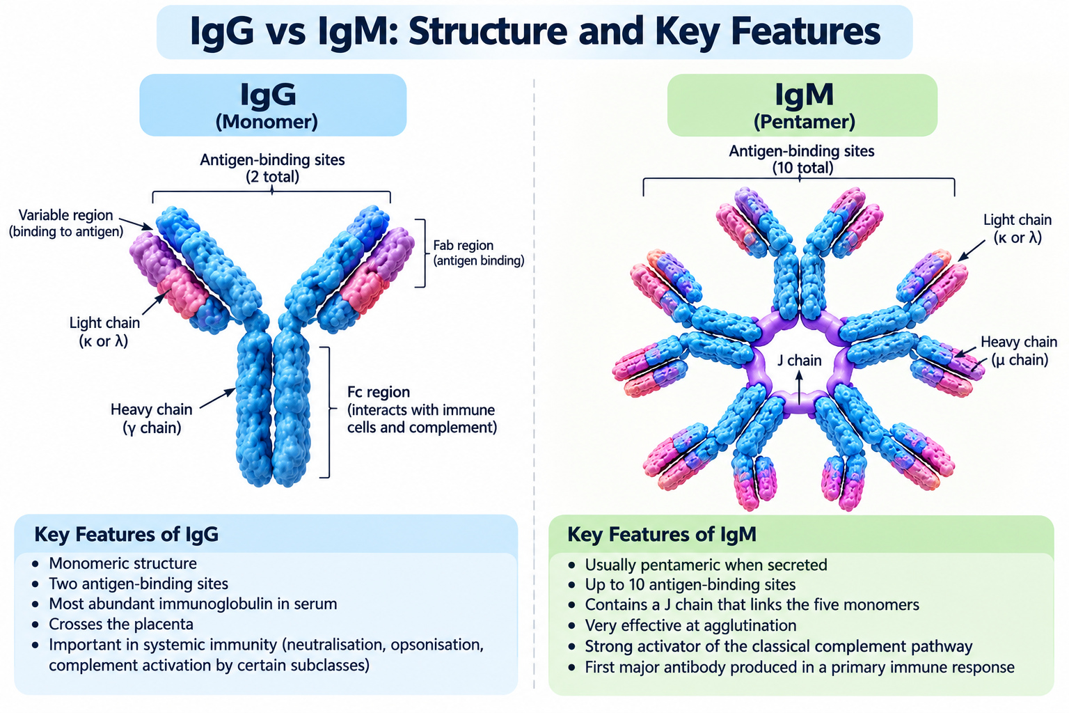
Secreted IgM is usually a pentamer, consisting of five antibody units joined together. Each unit contains two mu (μ) heavy chains and two light chains. The pentamer is stabilised by disulfide bonds and commonly includes a joining (J) chain that helps its assembly and secretion.
A typical IgM pentamer has up to 10 antigen-binding sites. However, the number of sites that can bind antigen simultaneously depends on the size, shape and arrangement of the antigen and on the accessibility of the binding sites.
The major structural features of IgM include:
- Mu heavy chains: Determine its immunoglobulin class.
- Pentameric arrangement: Gives secreted IgM a large molecular size and multiple antigen-binding sites.
- J chain: Participates in the assembly of polymeric IgM and contributes to its interaction with certain transport systems.
- High functional avidity: Multiple binding interactions can make IgM effective at binding repetitive structures on microbial surfaces.
Although IgM has multiple binding sites, its individual antigen-binding sites are not necessarily of higher affinity than those of mature IgG antibodies. Its strong overall binding often results from avidity, the combined strength of multiple antigen–antibody interactions.
Membrane-Bound and Secreted IgM
IgM occurs in two important forms.
Membrane-bound IgM: Present on the surface of mature naïve B lymphocytes, where it functions as part of the B-cell receptor. It is monomeric and helps the B cell recognise a specific antigen.
Secreted IgM: Released by antibody-secreting cells, especially during early immune responses. It is usually pentameric and functions in the extracellular environment.
This distinction is important because the statement that IgM is pentameric applies primarily to its secreted form, not to its membrane-bound B-cell receptor form.
Distribution of IgM
IgM is present mainly in blood plasma and lymph. Because of its relatively large molecular size, it does not distribute into tissues as readily as monomeric IgG.
IgM is also expressed on the surface of naïve B cells, often together with IgD. These membrane-bound immunoglobulins help B lymphocytes detect antigens and initiate an adaptive immune response.
In addition to newly produced IgM during infection, the body contains naturally occurring IgM antibodies. Some of these recognise common microbial structures or altered self-components and can contribute to early immune defence.
Major Functions of IgM
A. First Antibody in a Primary Immune Response
IgM is generally the first major antibody class secreted when naïve B cells respond to a new antigen. This makes it an important component of early humoral immunity.
As the response develops, activated B cells may undergo class-switch recombination and produce other classes, such as IgG, IgA or IgE, depending on the antigen and immune signals.
However, IgM is not restricted to primary responses. It can also be produced during later immune responses and by B-cell populations that maintain IgM production.
B. Agglutination of Antigens
Because secreted IgM has multiple antigen-binding sites, it is particularly effective at agglutination, the process of clumping cells or particles together.
For example, IgM can bind structures on the surfaces of bacteria or red blood cells and cross-link them into aggregates. This can facilitate their recognition and removal by immune mechanisms.
Agglutination is also important in laboratory tests, including some blood-grouping procedures.
C. Activation of the Classical Complement Pathway
One of the most important functions of IgM is its ability to activate the classical complement pathway after binding to an appropriate antigenic surface.
When IgM binds its target, its structure changes in a way that allows complement component C1q to interact with the antibody. This initiates a cascade of complement reactions that can:
- Promote inflammation.
- Enhance phagocytosis through complement-mediated opsonisation.
- Contribute to the destruction of susceptible target cells through the membrane attack complex.
A single antigen-bound IgM pentamer can be highly effective at initiating this pathway, although activation depends on the antibody and target surface.
D. Protection Against Blood-Borne Microorganisms
IgM is well suited to defence in the bloodstream because it circulates in plasma and binds effectively to many repetitive microbial surface structures.
By promoting agglutination and complement activation, IgM helps the immune system contain and eliminate certain bacteria and other pathogens before the adaptive response becomes fully developed.
E. Antigen Recognition by B Lymphocytes
Membrane-bound IgM is a major antigen-recognition molecule on mature naïve B cells. When a compatible antigen binds and appropriately cross-links B-cell receptors, it can initiate signalling that contributes to B-cell activation.
Further activation generally requires additional signals, which vary according to the antigen and the type of immune response.
IgM in Clinical Diagnosis
IgM tests are frequently used in the investigation of infectious diseases. The detection of pathogen-specific IgM may indicate a recent or current immune response to an infection, depending on the disease and test used.
However, the presence of IgM does not always prove a recent infection. IgM can persist after infection, produce false-positive results or react with related antigens. Conversely, an early test may be negative before sufficient antibody has developed.
For this reason, IgM results should be interpreted alongside symptoms, exposure history, the timing of testing and other diagnostic findings. In some infections, direct pathogen detection or additional antibody testing is needed to establish the diagnosis.
Difference Between IgM and IgG
| Feature | IgM | IgG |
|---|---|---|
| Heavy chain | Mu (μ) | Gamma (γ) |
| Secreted form | Usually pentameric | Monomeric |
| Antigen-binding sites | Up to 10 in a typical pentamer | 2 per molecule |
| Role in primary response | Often the first major class secreted | Usually develops after class switching, though timing varies |
| Complement activation | Particularly effective | Depends on the subclass and antigen-bound arrangement |
| Placental transfer | Does not normally cross the placenta in significant amounts | Crosses the placenta |
| Main distribution | Blood and lymph | Blood, extracellular fluid and tissues |
IgM is a major antibody of early immune defence. Its pentameric secreted form gives it high avidity for many antigens, strong agglutinating activity and exceptional ability to initiate the classical complement pathway. Its membrane-bound form also plays an essential role in antigen recognition by naïve B lymphocytes.
IgA: Structure, Distribution and Functions
Immunoglobulin A (IgA) is the principal antibody involved in protecting mucosal surfaces, including those lining the respiratory, gastrointestinal and genitourinary tracts. It is also present in saliva, tears and breast milk, where it helps prevent microorganisms and their products from attaching to and entering body tissues.
IgA is particularly important because many pathogens first encounter the body at mucosal surfaces. By acting at these entry points, IgA provides an important line of antibody-mediated defence while helping maintain a balanced relationship between the immune system and beneficial microorganisms.
Structure of IgA
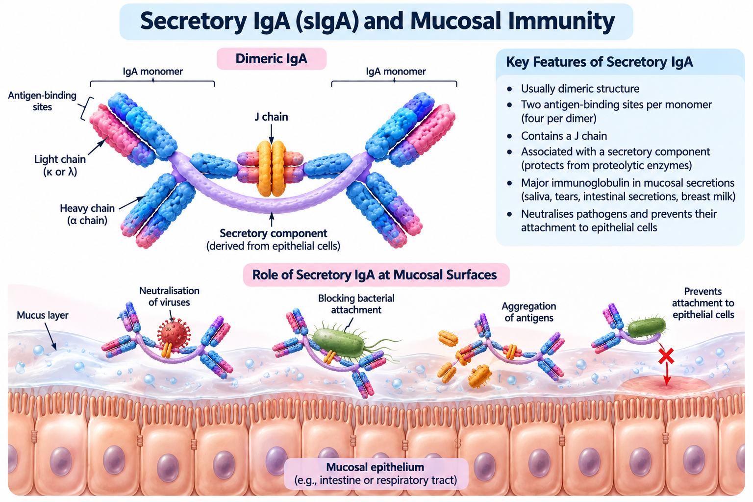
IgA contains alpha (α) heavy chains and occurs in two major structural forms: monomeric IgA and dimeric IgA.
Monomeric IgA consists of one antibody unit containing two heavy chains and two light chains. It is the predominant form of IgA in blood serum.
Dimeric IgA consists of two antibody units joined by a joining (J) chain. It is common in mucosal secretions and forms the basis of secretory IgA after transport across epithelial cells.
Secretory IgA contains an additional component called the secretory component, which is derived from the polymeric immunoglobulin receptor (pIgR) during transport through epithelial cells. This component helps protect IgA against degradation by enzymes present in mucosal secretions.
Important structural features include:
- Alpha heavy chains: Determine the IgA class.
- Two antigen-binding sites per monomer: Permit specific recognition of antigens.
- J chain: Helps join IgA units in polymeric forms and supports their transport across mucosal epithelial cells.
- Secretory component: Helps stabilise secretory IgA in mucosal environments.
The dimeric form has more antigen-binding sites than the monomeric form, allowing it to bind and cross-link multiple antigenic structures.
Distribution of IgA
IgA is distributed in both blood and mucosal secretions, but its form and role vary according to the location.
A. Blood Serum
In serum, IgA is found mainly as a monomer. It contributes to antibody-mediated immune defence in the circulation and tissues, although it is less abundant in serum than IgG.
B. Saliva and Tears
IgA is present in saliva and tears, where it helps protect the oral cavity and eyes. It can bind microorganisms and other antigens, reducing their ability to attach to epithelial surfaces.
C. Respiratory and Gastrointestinal Tracts
Secretory IgA is abundant in secretions covering mucosal surfaces. It contributes to protection in the respiratory tract and intestine, where pathogens may otherwise gain access to underlying tissues.
D. Breast Milk
Breast milk contains substantial amounts of secretory IgA. This antibody helps protect an infant’s intestinal mucosa by binding certain microorganisms and antigens. Its protective effects are particularly important during early infancy.
Major Functions of IgA
A. Protection of Mucosal Surfaces
The primary function of IgA is to protect mucosal surfaces from infection. It binds pathogens and antigens in mucus and other secretions, helping prevent their attachment to epithelial cells.
This mechanism is especially important in the respiratory and gastrointestinal tracts, where the body is continually exposed to microorganisms and environmental antigens.
B. Neutralisation of Pathogens and Toxins
IgA can neutralise viruses, bacterial toxins and other harmful substances. By binding to them, it may prevent their interaction with host cells.
Neutralisation by secretory IgA can occur at mucosal surfaces before pathogens enter the body’s tissues. This helps contain infection at its point of entry.
C. Prevention of Microbial Attachment
Many pathogens initiate infection by attaching to epithelial cells. Secretory IgA can bind microbial surface molecules and interfere with this attachment.
By reducing adherence, IgA helps prevent colonisation and supports the removal of microbes through mucus movement and other normal clearance mechanisms.
D. Immune Exclusion
Immune exclusion is the process by which antibodies, especially secretory IgA, bind antigens in mucosal secretions and help prevent their entry into underlying tissues.
IgA can trap microorganisms and antigens in mucus, cross-link them and facilitate their removal. This protective process limits exposure of epithelial tissues to potentially harmful agents.
E. Protection of the Intestinal Microbiota
The intestinal tract contains a large community of beneficial microorganisms known as the gut microbiota. IgA helps regulate interactions between these microorganisms and the host.
By binding certain microbial antigens, IgA can limit excessive contact with the intestinal epithelium and help maintain mucosal balance. Its effects depend on the microbial species, the antibody response and the local environment.
Importantly, IgA does not simply eliminate all intestinal microorganisms. It can also help the host coexist with beneficial microbes.
F. Passive Protection of Infants
Secretory IgA in breast milk provides mucosal protection to nursing infants. When consumed, it helps protect the infant’s gastrointestinal tract against certain pathogens and antigens.
This differs from the placental transfer of IgG. IgG provides systemic passive immunity before birth, whereas breast-milk IgA mainly supports protection at mucosal surfaces after birth.
IgA Subclasses
Humans have two IgA subclasses: IgA1 and IgA2. Both participate in mucosal and systemic immunity, but they differ in certain structural features and their distribution.
| Feature | IgA1 | IgA2 |
|---|---|---|
| Structural feature | Has a longer hinge region | Has a shorter hinge region |
| Distribution | Common in serum and several mucosal secretions | Particularly important in parts of the intestinal and other mucosal environments |
| Enzyme susceptibility | More susceptible to cleavage by certain bacterial IgA proteases | More resistant to many IgA1-specific bacterial proteases |
| General role | Contributes to antibody-mediated protection | Contributes to mucosal protection, including in environments containing IgA-degrading bacteria |
These differences help explain why the two subclasses are distributed differently across body compartments. Their functions overlap, and neither subclass is restricted to a single location.
Transport of IgA Across Epithelial Cells
Dimeric IgA produced by plasma cells in tissues beneath mucosal epithelium binds to the polymeric immunoglobulin receptor on the basolateral surface of epithelial cells. The receptor–IgA complex is transported through the cell and released at the mucosal surface.
During this process, part of the receptor remains attached to IgA as the secretory component. The resulting secretory IgA is better suited to function in environments where digestive enzymes and other proteases may degrade proteins.
This transport mechanism allows the immune system to deliver antibodies directly to mucosal surfaces without requiring large amounts of antibody to enter the bloodstream first.
Clinical and Diagnostic Importance of IgA
IgA measurements can help assess immune function and support the investigation of certain diseases. Low IgA levels may occur in selective IgA deficiency, a condition associated with an increased risk of some respiratory or gastrointestinal infections, although many affected individuals have no symptoms.
IgA is also important in the diagnosis of coeliac disease. Tests such as tissue transglutaminase IgA (tTG-IgA) are commonly used alongside assessment of total IgA, because IgA deficiency can make an IgA-based test falsely reassuring. Alternative testing strategies may be needed when IgA deficiency is present.
Elevated IgA can occur in several inflammatory, infectious and immune-related conditions. As with other immunoglobulin tests, results must be interpreted in the context of symptoms and other clinical findings.
IgA is the major antibody class associated with mucosal defence. Its secretory form protects surfaces such as the respiratory and intestinal tracts by neutralising pathogens, preventing microbial attachment and helping maintain a healthy relationship between the host and its microbiota.
IgE: Structure, Distribution and Functions
Immunoglobulin E (IgE) is an antibody class primarily involved in allergic reactions and immune defence against certain parasitic worms, particularly helminths. Although IgE is present in blood serum at very low concentrations compared with IgG, IgA and IgM, it can trigger powerful cellular responses when it binds to specific receptors on mast cells and basophils.
IgE plays a protective role in some immune responses, but inappropriate or excessive activation of IgE-associated pathways can contribute to allergic conditions such as allergic rhinitis, asthma and anaphylaxis.
Structure of IgE
IgE is a monomeric immunoglobulin composed of two identical heavy chains and two identical light chains. Its heavy chains are called epsilon (ε) chains.
Like other antibody classes, IgE has antigen-binding regions that recognise specific epitopes. However, its heavy-chain constant region differs structurally from those of other immunoglobulins. The epsilon heavy chain contains four constant domains and lacks the typical flexible hinge region found in IgG.
Important structural features of IgE include:
- Epsilon heavy chains: Determine the IgE class.
- Two antigen-binding sites: Allow the molecule to recognise specific epitopes.
- Four constant domains in each heavy chain: Contribute to the structure and receptor interactions of IgE.
- Fc region: Binds to specialised Fc receptors on immune cells.
- High-affinity receptor binding: Allows IgE to remain attached to mast cells and basophils even when free IgE concentrations in serum are low.
The Fc region is especially important because IgE usually exerts its major biological effects through receptor-bearing cells rather than by circulating freely in large quantities.
Distribution of IgE
IgE is found in very low concentrations in blood serum. A substantial proportion of its biological activity is associated with its binding to immune cells.
A. Mast Cells
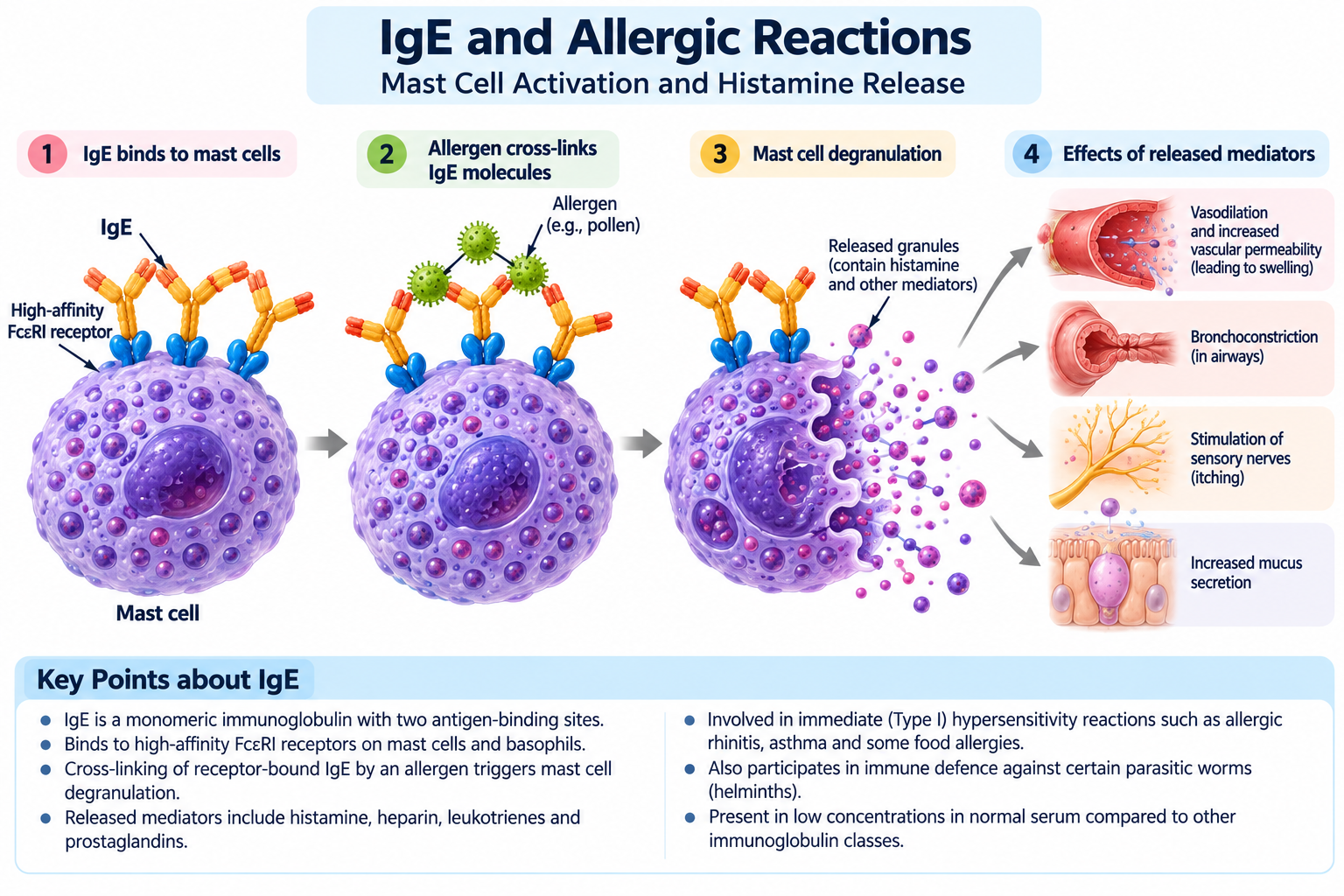
Mast cells are tissue-resident immune cells found in locations such as the skin, respiratory tract and gastrointestinal tract. They express high-affinity IgE receptors called FcεRI.
IgE molecules bind to these receptors and sensitise the mast cells to their corresponding antigens. If a suitable antigen later cross-links adjacent receptor-bound IgE molecules, the mast cells can become activated and release inflammatory mediators.
B. Basophils
Basophils are circulating white blood cells that also express high-affinity IgE receptors. When appropriately activated, they release substances that contribute to inflammation and allergic responses.
C. Tissues and Mucosal Sites
IgE-associated responses occur particularly at barrier sites exposed to environmental allergens and parasites. These include the skin, respiratory tract and gastrointestinal tract.
Major Functions of IgE
A. Immediate Hypersensitivity Reactions
The best-known function of IgE is its role in type I hypersensitivity, also called immediate hypersensitivity.
The process generally occurs in two stages:
1. Sensitisation: During an initial allergic response, the immune system produces allergen-specific IgE. This IgE binds to FcεRI receptors on mast cells and basophils.
2. Re-exposure: When the same allergen is encountered again, it may cross-link receptor-bound IgE molecules. This activates the cells and can trigger the release of inflammatory mediators.
The released substances can cause itching, swelling, mucus production, airway narrowing and other allergic symptoms. The severity depends on the allergen, the tissues involved and the extent of the immune response.
B. Release of Histamine and Other Mediators
Following activation, mast cells and basophils release histamine and other inflammatory mediators. These substances produce several physiological effects.
- Increased vascular permeability: Fluid moves from blood vessels into surrounding tissues, contributing to swelling.
- Vasodilation: Blood vessels widen, contributing to redness and warmth.
- Smooth-muscle contraction: In the airways, this may cause bronchoconstriction.
- Increased mucus secretion: This contributes to nasal congestion and other respiratory symptoms.
- Sensory nerve stimulation: This may cause itching and sneezing.
Other mediators released during allergic reactions contribute to prolonged inflammation and recruitment of additional immune cells.
C. Defence Against Helminths
IgE also participates in immune responses against certain parasitic worms, known as helminths.
In these responses, parasite-specific IgE may bind to the parasite and interact with immune cells bearing IgE receptors. Alongside other mechanisms involving eosinophils, mast cells and type 2 immune responses, these interactions can contribute to parasite defence.
However, the importance of IgE varies among parasites, and not every helminth infection is controlled primarily through IgE.
D. Recruitment and Activation of Immune Cells
IgE-associated responses can help recruit and activate cells involved in inflammation and parasite defence. Mast cells release mediators that influence nearby blood vessels and immune cells, while other components of type 2 immunity contribute to the overall response.
These reactions can be beneficial when directed against certain parasites but harmful when triggered excessively by harmless environmental substances.
Mechanism of IgE-Mediated Allergic Reactions
The mechanism can be summarised as follows:
- An allergen enters the body and stimulates an appropriate immune response.
- Allergen-specific IgE is produced by antibody-secreting cells.
- IgE binds to high-affinity FcεRI receptors on mast cells and basophils.
- On subsequent exposure, the allergen cross-links receptor-bound IgE.
- The cells become activated and release histamine and other mediators.
- The resulting inflammation produces allergic symptoms.
Not every exposure to an allergen produces a severe reaction. The outcome depends on the degree of sensitisation, the amount and route of exposure, and individual susceptibility.
IgE and Anaphylaxis
Anaphylaxis is a potentially life-threatening systemic reaction that can develop rapidly after exposure to a triggering substance. IgE-mediated mast-cell and basophil activation is an important mechanism in many cases, although not all anaphylactic reactions follow the same pathway.
Symptoms may include difficulty breathing, swelling of the tongue or throat, widespread hives, dizziness and a sudden fall in blood pressure.
Suspected anaphylaxis is a medical emergency. Intramuscular adrenaline (epinephrine) is the first-line treatment, and emergency medical assistance is required. Antihistamines do not replace adrenaline in treating anaphylaxis.
Clinical and Diagnostic Importance of IgE
IgE testing can help investigate suspected allergic disease. Two commonly used approaches are:
- Total IgE testing: Measures the overall concentration of IgE in serum.
- Allergen-specific IgE testing: Measures IgE directed against a particular allergen or allergen extract.
An elevated total IgE level does not, by itself, prove that a person has an allergy. Levels may also be elevated in certain parasitic infections and other conditions, while some people with allergic disease have normal total IgE levels.
Allergen-specific IgE results must also be interpreted alongside clinical history and symptoms. A positive result indicates sensitisation but does not necessarily mean that exposure will cause clinical symptoms.
Difference Between IgE and Other Immunoglobulins
IgE differs from the other major antibody classes in its specialised receptor interactions and its role in immediate hypersensitivity.
| Feature | IgE | IgG |
|---|---|---|
| Heavy chain | Epsilon (ε) | Gamma (γ) |
| Main structural form | Monomer | Monomer |
| Serum concentration | Very low | Relatively high |
| Major receptor-associated role | Activates mast cells and basophils through FcεRI | Supports functions such as phagocytosis and cellular cytotoxicity through Fc receptors |
| Major immune roles | Allergic reactions and defence against certain helminths | Neutralisation, opsonisation and other systemic defence mechanisms |
| Placental transfer | Does not normally cross the placenta in significant amounts | Crosses the placenta |
IgE is a specialised antibody class that binds strongly to receptors on mast cells and basophils. It contributes to defence against certain parasitic worms but is also central to many immediate allergic reactions, including potentially life-threatening anaphylaxis.
IgD: Structure, Distribution and Functions
Immunoglobulin D (IgD) is one of the five major classes of immunoglobulins. It is found primarily on the surface of mature naïve B lymphocytes, where it functions as an antigen receptor and participates in B-cell activation. Although IgD is present at relatively low concentrations in blood serum, it has an important role in the development and regulation of adaptive humoral immunity.
Compared with IgG, IgM and IgA, the functions of IgD are less extensively understood. However, research has established its importance as a B-cell surface receptor and suggests additional roles in immune responses at respiratory mucosal surfaces.
Structure of IgD
IgD is a monomeric immunoglobulin consisting of two identical heavy chains and two identical light chains. Its heavy chains are called delta (δ) chains.
Like other monomeric antibodies, IgD contains two antigen-binding sites. Its heavy-chain constant region has three constant domains and an extended hinge region, which provides flexibility for antigen recognition.
The main structural features of IgD include:
- Delta heavy chains: Determine the immunoglobulin class.
- Two light chains: Participate in the formation of the antigen-binding regions.
- Two antigen-binding sites: Recognise specific epitopes.
- Extended hinge region: Provides flexibility to the antibody molecule.
- Membrane-bound form: Functions as a receptor on B lymphocytes.
- Secreted form: Occurs in small amounts and may contribute to immune responses outside the B-cell surface.
The membrane-bound and secreted forms arise through alternative processing of immunoglobulin heavy-chain transcripts. Their different forms allow IgD to participate in both cell-surface antigen recognition and extracellular immune activity.
Distribution of IgD
IgD is distributed mainly in two forms.
A. On the Surface of B Lymphocytes
The major location of IgD is the surface of mature naïve B cells. These cells commonly express both IgM and IgD as antigen receptors with the same antigen specificity.
Although the two receptors recognise the same antigen, they differ in their constant regions and associated signalling properties. Their expression helps B cells detect antigens and respond appropriately to immune signals.
B. In Blood Serum
Small amounts of secreted IgD are present in blood serum. Its concentration is much lower than that of IgG and several other major immunoglobulin classes.
Because serum IgD is relatively scarce, its precise contribution to general circulating antibody protection is less clearly established than that of IgG, IgM or IgA.
C. At Respiratory Mucosal Surfaces
IgD-producing B cells and secreted IgD have been studied in relation to the upper respiratory tract. IgD may interact with immune cells and microbial antigens at these sites.
However, these proposed and observed activities should not be interpreted as making IgD the principal mucosal antibody. IgA remains the major immunoglobulin associated with mucosal secretions.
Major Functions of IgD
A. Antigen Recognition by B Cells
The best-established function of IgD is its role as an antigen receptor on mature naïve B lymphocytes.
When a B-cell receptor recognises a compatible antigen, it can initiate intracellular signalling. This signalling contributes to B-cell activation, although additional signals are generally required for a complete immune response.
Antigen recognition allows B cells to respond selectively to substances that match the specificity of their receptors.
B. Participation in B-Cell Activation
IgD works alongside IgM on the surface of mature naïve B cells. Together, these receptors help the cells recognise antigens and participate in the early stages of the adaptive immune response.
The activation and subsequent development of a B cell depend on several factors, including antigen properties, receptor signalling and signals from other immune cells. IgD is therefore one component of a broader regulatory system rather than an antibody that acts independently.
C. Regulation of B-Cell Responses
The expression of IgD is associated with the maturation and functional regulation of B lymphocytes. Differences in IgM and IgD expression can influence how B cells respond to antigen exposure and their surrounding environment.
The exact contribution of IgD to these regulatory processes varies with the B-cell population and the biological context.
D. Possible Contributions to Mucosal Immunity
Secreted IgD has been investigated for its ability to interact with respiratory microorganisms and immune cells. These interactions may contribute to local immune responses at mucosal surfaces.
Nevertheless, the precise protective role of IgD in humans remains an area of continuing research. Its contribution should not be overstated relative to the well-established roles of IgA in mucosal protection and IgG in systemic immunity.
IgD and IgM: Their Relationship on B Cells
IgD and IgM are particularly important because both can function as antigen receptors on mature naïve B lymphocytes.
| Feature | IgD | IgM |
|---|---|---|
| Heavy chain | Delta (δ) | Mu (μ) |
| Typical B-cell surface form | Monomer | Monomer |
| Secreted form | Present in small amounts | Usually pentameric |
| Expression on mature naïve B cells | Commonly expressed | Commonly expressed |
| Main recognised role | B-cell antigen recognition and regulation | B-cell antigen recognition and early antibody defence |
| Abundance in serum | Very low | Greater than IgD |
Despite their differences, membrane-bound IgD and IgM on the same naïve B cell generally recognise the same antigen because their variable regions share the same antigen specificity.
Clinical and Diagnostic Importance of IgD
IgD is not routinely measured in the same way as IgG, IgA and IgM in many standard assessments of immune function. However, it has clinical relevance in selected conditions.
For example, some rare plasma-cell disorders produce monoclonal IgD. In such cases, laboratory investigations may include specialised immunoglobulin measurements and protein studies to identify the abnormal antibody.
IgD has also been investigated in inflammatory and immune-mediated conditions, but its significance depends on the specific disease and available evidence. An isolated IgD measurement is generally insufficient to establish a diagnosis.
IgD is a minor circulating immunoglobulin with an important role on the surface of mature naïve B lymphocytes. Working alongside IgM, it participates in antigen recognition and B-cell activation, helping initiate the adaptive immune response.
Comparison of the Five Immunoglobulin Classes
The five immunoglobulin classes—IgG, IgM, IgA, IgE and IgD—share the basic antibody structure but differ in their heavy chains, molecular arrangements, distribution and biological functions. These differences allow the immune system to provide protection in the bloodstream, tissues and mucosal surfaces while coordinating specialised immune responses.
Understanding the differences between the five classes is important for studying humoral immunity, interpreting immunological tests and preparing for zoology examinations.
General Comparison of Immunoglobulins
| Feature | IgG | IgM | IgA | IgE | IgD |
|---|---|---|---|---|---|
| Heavy chain | Gamma (γ) | Mu (μ) | Alpha (α) | Epsilon (ε) | Delta (δ) |
| Main structural form when secreted | Monomer | Usually pentamer | Mainly monomer in serum; often dimer in secretions | Monomer | Monomer |
| Antigen-binding sites per typical secreted molecule | 2 | Up to 10 in a pentamer | 2 per monomer; commonly 4 in a dimer | 2 | 2 |
| Relative serum abundance | Highest | Second most abundant in typical adult serum | Lower than IgG and IgM | Very low | Very low |
| Major location | Blood and tissues | Blood and lymph | Mucosal secretions and blood | Mainly bound to mast cells and basophils | Mainly B-cell surfaces |
| Principal role | Systemic protection and neutralisation | Early antibody response and complement activation | Mucosal protection | Allergic responses and defence against certain parasites | B-cell antigen recognition |
| Placental transfer | Yes | No significant normal transfer | No significant normal transfer | No significant normal transfer | No significant normal transfer |
Note: Serum abundance varies with age, health and individual circumstances. The number of antigen-binding sites indicates the typical structural capacity of each form; not all sites necessarily bind antigen simultaneously.
Differences in Molecular Structure
The immunoglobulin class is determined by the constant region of its heavy chains. These structural differences influence how antibodies interact with immune cells, complement proteins and other biological systems.
- IgG is monomeric and readily distributes from blood into extracellular tissues.
- IgM is usually pentameric when secreted, giving it multiple antigen-binding sites and high overall avidity for many antigens.
- IgA occurs mainly as a monomer in serum and commonly as a dimer in mucosal secretions.
- IgE is monomeric and has a constant region adapted for binding to specialised Fc receptors.
- IgD is monomeric and is particularly important as a membrane-bound receptor on mature naïve B lymphocytes.
The different structures are not merely physical variations. They help determine where each antibody acts and which immune mechanisms it can efficiently initiate.
Differences in Distribution
Immunoglobulins are distributed according to their molecular properties and biological roles.
IgG: Its relatively small monomeric structure allows it to circulate in blood and enter tissues. It is also transported across the placenta, providing passive immunity to the foetus.
IgM: Its large pentameric structure means that secreted IgM is concentrated mainly in blood and lymph. Its membrane-bound form occurs on B cells as an antigen receptor.
IgA: Its secretory form is adapted to mucosal environments, including the respiratory and gastrointestinal tracts. It is also present in saliva, tears and breast milk.
IgE: Although found in low concentrations in serum, IgE binds strongly to high-affinity receptors on mast cells and basophils. These cell-associated antibodies participate in allergic and other immune responses.
IgD: It is found mainly on the surface of mature naïve B cells, where it contributes to antigen recognition and B-cell activation.
Differences in Biological Functions
Each immunoglobulin class contributes to immune defence in a characteristic way.
| Immunoglobulin | Important functions |
|---|---|
| IgG | Neutralisation, opsonisation, complement activation by certain subclasses, antibody-dependent cellular cytotoxicity and placental transfer |
| IgM | Early antibody defence, agglutination, classical complement activation and B-cell antigen recognition |
| IgA | Neutralisation at mucosal surfaces, prevention of microbial attachment and protection of the intestinal and respiratory tracts |
| IgE | Immediate hypersensitivity reactions, mast-cell activation and responses against certain helminths |
| IgD | Antigen recognition and regulation of responses by mature naïve B lymphocytes |
These functions overlap in some areas. For example, both IgG and IgA can neutralise pathogens, but IgG is especially important in systemic protection while IgA is specialised for mucosal defence.
Which Immunoglobulin Is Produced First?
IgM is generally the first major antibody class secreted during a primary response to a new antigen. It can provide early protection through agglutination and complement activation.
As the immune response develops, activated B cells may undergo class-switch recombination and produce IgG, IgA or IgE, depending on the signals they receive. The resulting antibody classes perform specialised functions while retaining antigen specificity when the variable region remains unchanged.
IgM is not limited to the first encounter, and IgG is not restricted to secondary responses. Both classes may be present at different stages of an immune response.
Which Immunoglobulin Provides Long-Term Protection?
IgG is particularly important in long-term antibody-mediated protection because it can persist in circulation and be produced by long-lived plasma cells. Memory B cells also support faster antibody production after subsequent exposure to a previously encountered antigen.
However, long-term protection is not provided by IgG alone. Memory B cells can produce different antibody classes, and mucosal immunity may involve IgA. The type of protection depends on the pathogen, the site of infection and the immune response generated.
- IgG: Most abundant immunoglobulin in adult human serum and the principal class transported across the placenta.
- IgM: Usually the first major antibody class secreted in a primary immune response; secreted IgM is typically pentameric.
- IgA: Major antibody class in mucosal secretions; secretory IgA is commonly dimeric.
- IgE: Associated with immediate allergic reactions and defence against certain parasitic worms.
- IgD: Mainly functions as an antigen receptor on mature naïve B lymphocytes.
These distinctions provide a useful foundation for understanding the roles of immunoglobulins in humoral immunity and their clinical significance.
Primary and Secondary Immune Responses: Roles of Antibody Classes
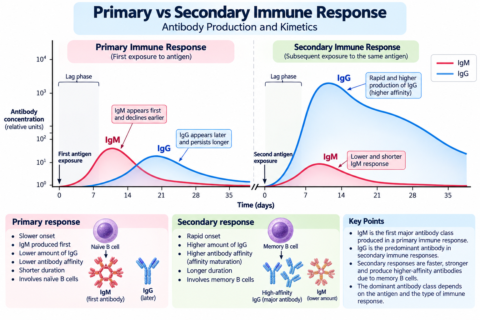
The production of immunoglobulins changes as the immune system encounters an antigen for the first time and then encounters it again. These responses are called the primary immune response and the secondary immune response, respectively.
The two responses differ in their speed, antibody concentration, antibody affinity and contribution from memory B cells. IgM is generally the first major antibody class secreted during a primary response, whereas IgG often becomes prominent during later stages and secondary responses. IgA and IgE may also be produced when appropriate immune signals direct B cells towards these classes.
B lymphocytes and other immune cells coordinate antibody production and immune memory. Read our guide to the cells of the immune system to understand their individual roles.
Primary Immune Response
The primary immune response occurs when the immune system encounters a particular antigen for the first time.
During this response, naïve B lymphocytes that recognise the antigen become activated with the help of appropriate signals. They proliferate and differentiate into antibody-secreting plasma cells and memory B cells.
The response generally develops through the following stages:
- Antigen recognition: A specific B cell recognises the antigen through its B-cell receptor.
- B-cell activation: Additional signals stimulate the B cell to become activated. For many protein antigens, these signals involve helper T cells.
- Clonal expansion: The activated B cell proliferates, producing a population of cells with the same antigen specificity.
- Initial antibody production: IgM is generally the first major class secreted.
- Class switching and affinity maturation: Under suitable conditions, some B cells switch to producing other immunoglobulin classes and undergo processes that can improve antibody affinity.
- Memory formation: Some activated B cells become memory B cells, which can respond more rapidly to future exposure.
The timing and magnitude of these stages depend on factors such as antigen type, dose, route of entry and the individual’s immune status.
Role of IgM in the Primary Response
IgM is particularly important during the early phase of the primary immune response. Its pentameric secreted form binds multiple antigenic sites and can efficiently activate the classical complement pathway.
This helps provide early antibody-mediated protection before substantial quantities of other antibody classes have accumulated.
However, IgM production does not stop when IgG or other classes appear. Some B cells continue producing IgM, and IgM can also be involved in responses to previously encountered antigens.
Role of Other Immunoglobulin Classes
As the immune response develops, different B-cell populations may produce antibodies adapted to particular locations and functions.
- IgG: Often becomes prominent in the later stages of a primary response and is frequently important in secondary responses. It supports neutralisation, opsonisation and other systemic defence mechanisms.
- IgA: Contributes to protection at mucosal surfaces when the response involves sites such as the respiratory or gastrointestinal tract.
- IgE: May be produced during immune responses directed towards certain parasites. It also participates in allergic sensitisation and immediate hypersensitivity.
- IgD: Functions mainly as a receptor on mature naïve B cells rather than as a major secreted antibody in the conventional primary antibody response.
Class switching changes the constant region of the heavy chain while generally preserving the antigen specificity encoded by the variable region. Consequently, antibodies of different classes can recognise the same antigen but carry out different biological functions.
Secondary Immune Response
The secondary immune response occurs when the immune system encounters the same antigen again after an earlier response has generated immunological memory.
Memory B cells can recognise the previously encountered antigen and respond more rapidly than naïve B cells. Depending on the antigen and the memory B-cell population, this response may produce antibodies in a shorter period, reach a higher antibody concentration and generate antibodies with greater average affinity.
The secondary response commonly contains substantial amounts of IgG, although IgA or other classes may also be important depending on the location and nature of the original immune response.
Major characteristics include:
- Shorter lag period: Antibody production often begins more quickly.
- Greater antibody concentration: The response can produce more antigen-specific antibody.
- Improved average affinity: Selection during affinity maturation can result in memory responses dominated by higher-affinity antibodies.
- Class-switched antibodies: IgG and, where appropriate, IgA or IgE may contribute significantly.
- Memory B-cell participation: Previously generated antigen-specific cells contribute to the enhanced response.
The secondary response is not necessarily identical for every antigen. Its strength and antibody composition depend on the type of antigen, the immune history of the individual and the nature of the memory cells produced.
Primary vs Secondary Immune Response
| Feature | Primary immune response | Secondary immune response |
|---|---|---|
| Exposure | First encounter with a particular antigen | Subsequent encounter with the same antigen |
| Main B-cell population involved | Naïve B cells | Memory B cells, alongside other responding B cells |
| Time to develop | Generally slower | Generally faster |
| Early major antibody class | Usually IgM | Often a substantial response from class-switched antibodies, especially IgG |
| Antibody concentration | Usually lower | Often higher |
| Average antibody affinity | Initially lower, with affinity maturation occurring during the response | Often higher because of prior selection and maturation |
| Memory-cell formation | Memory cells are generated | Existing memory cells contribute to the response |
These are general patterns rather than absolute rules. For example, IgM can also appear during secondary responses, and some primary responses generate class-switched antibodies relatively early.
Importance in Vaccination
Vaccination uses immunological memory to help the body respond effectively to future exposure to a pathogen.
A vaccine presents an antigen or instructions for producing an antigen in a controlled manner. This stimulates an immune response that may generate antigen-specific antibodies, memory B cells and, depending on the vaccine, other forms of immune memory.
A subsequent vaccine dose or exposure can stimulate a stronger or more rapid response in many circumstances. Booster doses are designed to strengthen or restore protection when needed.
The antibody class produced depends on the vaccine, antigen, route of administration and immune signals generated. Many vaccines induce important IgG responses, while vaccines delivered through mucosal routes may also stimulate mucosal IgA responses.
Why Are Different Immunoglobulin Classes Necessary?
The immune system requires more than one antibody class because pathogens enter the body through different routes and occupy different biological environments.
- IgM supports early defence in blood and lymph.
- IgG provides widespread systemic protection and can cross the placenta.
- IgA protects mucosal surfaces, where many infections begin.
- IgE participates in specialised responses against certain parasites and in immediate allergic reactions.
- IgD contributes to antigen recognition by mature naïve B lymphocytes.
Together, these classes allow antibody-mediated immunity to respond to diverse threats rather than relying on a single antibody type.
The primary immune response establishes antigen-specific immunity and immunological memory, while the secondary response can provide faster and more effective protection. IgM is generally the first major class secreted during a primary response, whereas IgG often plays a prominent role in later and secondary responses. IgA and IgE provide additional specialised protection, while IgD contributes mainly to B-cell antigen recognition.
Clinical and Diagnostic Importance of Immunoglobulins
Immunoglobulins are important not only for protecting the body against pathogens but also for diagnosing and monitoring several diseases. Their concentrations, subclasses and antigen-specific responses can provide useful information about immune function, previous exposure to infections, allergic sensitisation and certain immune-related disorders.
Laboratory investigations may measure total immunoglobulin levels, detect antibodies against particular antigens or identify abnormal immunoglobulin proteins. The results must be interpreted alongside clinical symptoms, medical history and other investigations because an antibody result alone rarely establishes a diagnosis.
The development of antibody responses depends on the coordinated activity of immune cells and the organs where they develop and interact. Learn more about these structures in our article on lymphoid organs.
Measurement of Immunoglobulin Levels
Quantitative immunoglobulin tests measure the concentration of major antibody classes in blood serum. Commonly measured classes include IgG, IgA and IgM. IgE and IgD may be measured when there is a specific clinical reason.
These tests can help investigate suspected immune deficiencies, recurrent infections and certain inflammatory or blood disorders.
- Low immunoglobulin levels: May indicate reduced antibody production, an immunodeficiency condition, protein loss or another underlying problem.
- Elevated immunoglobulin levels: May occur in chronic infections, inflammatory conditions, autoimmune diseases and some blood disorders.
- Abnormal patterns across several classes: May help clinicians distinguish different types of immune dysfunction.
Results are interpreted using age-appropriate reference ranges and the patient’s clinical circumstances. A low or high result does not necessarily mean that the immune system is globally weak or overactive.
Immunoglobulins in the Diagnosis of Infections
Antigen-specific antibodies can help determine whether the immune system has responded to a particular pathogen. Depending on the infection, testing may detect IgM, IgG or both.
IgM antibodies: May indicate a relatively recent immune response, but they can persist after infection or produce false-positive results. Their presence does not automatically establish an acute infection.
IgG antibodies: May indicate previous exposure, vaccination or an established immune response. In some infections, the appearance or change in IgG levels between appropriately timed samples provides useful diagnostic information.
The interpretation varies by pathogen and test. In certain situations, direct detection methods—such as polymerase chain reaction (PCR), antigen testing or culture—are more useful for determining whether a pathogen is currently present.
Immunoglobulins and Vaccination
Vaccines stimulate immune responses that may generate antigen-specific antibodies and immunological memory. Antibody measurements can sometimes be used to assess the response to vaccination or help evaluate suspected antibody deficiencies.
For example, clinicians may assess antibody responses to selected vaccine antigens when investigating whether a person produces adequate specific antibodies.
However, the presence of a measurable antibody does not always guarantee complete protection, and the absence of a particular antibody from a test does not necessarily mean that all immune memory is absent. The protective threshold and usefulness of antibody testing depend on the pathogen, vaccine and assay.
Immunoglobulins in Allergic Diseases
IgE is particularly important in the investigation of immediate allergic reactions.
Two common investigations are:
- Total IgE: Measures the overall concentration of IgE in serum.
- Allergen-specific IgE: Detects IgE directed against a particular allergen or allergen extract.
These tests can support the assessment of suspected allergic conditions, including allergic rhinitis and some forms of allergic asthma.
Nevertheless, an elevated total IgE level does not prove that a person has a specific allergy. Similarly, a positive allergen-specific IgE result indicates sensitisation but does not always mean that exposure causes symptoms. Clinical history and, where appropriate, other diagnostic assessments are essential.
Immunoglobulins in Autoimmune Diseases
Autoimmune diseases occur when immune responses target the body’s own tissues. Some autoimmune conditions are associated with antibodies directed against self-antigens, known as autoantibodies.
Examples include antinuclear antibodies (ANA) in the investigation of certain systemic autoimmune diseases and antibodies against specific tissue components in other conditions.
Autoantibodies are not all immunoglobulins with the same biological effect, and their clinical meaning depends on the antigen they recognise, the assay used and the patient’s symptoms. Some autoantibodies may also occur in people without a diagnosed autoimmune disease.
Consequently, autoimmune diagnosis usually requires a combination of clinical assessment, laboratory findings and sometimes imaging or tissue examination.
Immunoglobulins in Immunodeficiency Disorders
Immunodeficiency conditions can affect the production, function or regulation of antibodies. Some people have reduced concentrations of one or more immunoglobulin classes, while others have normal total levels but inadequate responses to specific antigens.
Examples include:
- Selective IgA deficiency: Characterised by markedly reduced IgA with other major immunoglobulin classes generally within expected ranges, after relevant causes have been excluded.
- Common variable immunodeficiency (CVID): A heterogeneous condition commonly associated with reduced IgG and reduced IgA and/or IgM, impaired specific antibody responses and a compatible clinical history.
- X-linked agammaglobulinaemia: A condition involving a profound reduction in mature B cells and antibody production.
The clinical effects vary. Some affected individuals experience recurrent respiratory or gastrointestinal infections, while others may have milder symptoms. Diagnosis requires appropriate laboratory evaluation and exclusion of other causes.
Immunoglobulins in Coeliac Disease
Coeliac disease is an immune-mediated condition triggered by gluten in genetically susceptible individuals. Antibody testing plays an important role in its investigation.
A commonly used initial test is tissue transglutaminase IgA (tTG-IgA), often accompanied by measurement of total IgA. The total IgA measurement is important because IgA deficiency can cause an IgA-based test to appear negative even when coeliac disease is present.
When IgA deficiency is identified or suspected, clinicians may use appropriate IgG-based tests, such as IgG antibodies against deamidated gliadin peptides, depending on the clinical context.
Testing should generally be performed while the person is consuming gluten, unless a clinician advises otherwise. Test results must be interpreted within the relevant diagnostic pathway.
Monoclonal Immunoglobulins and Blood Disorders
Immunoglobulins are produced by populations of antibody-secreting cells. In some plasma-cell disorders, a single abnormal cell population produces large amounts of one immunoglobulin or immunoglobulin fragment. This is called a monoclonal protein or paraprotein.
Monoclonal proteins may be detected during investigations for conditions such as multiple myeloma and other related plasma-cell disorders.
Laboratory methods may include serum protein electrophoresis, immunofixation and serum free light-chain testing. These investigations help characterise abnormal proteins, but an abnormal result does not by itself establish a specific malignancy.
Further evaluation is guided by the clinical presentation and other laboratory or imaging findings.
Summary of Clinical Applications
| Clinical application | Relevant immunoglobulin or test | Main purpose |
|---|---|---|
| Investigation of antibody deficiency | IgG, IgA, IgM and specific antibody responses | Assess antibody production and function |
| Investigation of certain infections | Pathogen-specific IgM and IgG | Support assessment of exposure or immune response |
| Allergy assessment | Total and allergen-specific IgE | Investigate suspected IgE-mediated sensitisation |
| Coeliac disease testing | tTG-IgA and total IgA | Support diagnosis and identify potential IgA-related testing limitations |
| Autoimmune disease investigation | Disease-associated autoantibodies | Support evaluation of suspected autoimmune conditions |
| Plasma-cell disorder investigation | Protein electrophoresis, immunofixation and related tests | Detect and characterise abnormal monoclonal proteins |
Immunoglobulin testing provides valuable information about immune function and can support the investigation of infections, allergies, autoimmune diseases, immunodeficiencies and plasma-cell disorders. Its greatest value comes from interpreting the results in the appropriate clinical context rather than relying on a single antibody measurement.
FAQs
What are the five types of immunoglobulins?
Which immunoglobulin is most abundant in human blood serum?
Which immunoglobulin is produced first during a primary immune response?
Which immunoglobulin crosses the placenta?
Which immunoglobulin is mainly responsible for mucosal immunity?
Which immunoglobulin is associated with allergic reactions?
Which immunoglobulin is mainly found on the surface of mature naïve B lymphocytes?
Why is IgM called a pentamer?
What is the difference between IgA1 and IgA2?
What is the difference between IgG and IgM?
Can IgM indicate a recent infection?
What is class-switch recombination?
Which immunoglobulin is involved in defence against parasitic worms?
Which immunoglobulin is most important in secondary immune responses?
Are immunoglobulins and antibodies the same thing?
Key Takeaways Types of Immunoglobulins
- Immunoglobulins are glycoproteins produced by plasma cells and are essential components of humoral immunity.
- The five major immunoglobulin classes are IgG, IgM, IgA, IgE and IgD.
- The immunoglobulin class is determined by the constant region of its heavy chain.
- The heavy chains of IgG, IgM, IgA, IgE and IgD are gamma (γ), mu (μ), alpha (α), epsilon (ε) and delta (δ), respectively.
- IgG is the most abundant immunoglobulin in normal adult human serum and is the principal class transported across the placenta.
- IgM is generally the first major antibody class secreted during a primary immune response.
- Secreted IgM is usually pentameric and can have up to 10 antigen-binding sites.
- Membrane-bound IgM is monomeric and functions as a B-cell receptor.
- IgA is the major immunoglobulin associated with mucosal secretions, including saliva, tears and intestinal secretions.
- Secretory IgA commonly occurs as a dimer containing a J chain and a secretory component.
- Human IgA has two subclasses: IgA1 and IgA2.
- IgE participates in immediate allergic reactions and immune defence against certain parasitic worms.
- IgE binds to high-affinity FcεRI receptors on mast cells and basophils.
- IgD is found mainly on mature naïve B lymphocytes, where it functions as an antigen receptor.
- IgG has four human subclasses: IgG1, IgG2, IgG3 and IgG4.
- Class-switch recombination changes the immunoglobulin heavy-chain constant region while generally retaining antigen specificity.
- During secondary immune responses, memory B cells can produce antibodies more rapidly and often generate higher-affinity responses.
- Immunoglobulin tests support the investigation of infections, allergies, immunodeficiencies, autoimmune conditions and certain blood disorders.
Conclusion
The five types of immunoglobulins—IgG, IgM, IgA, IgE and IgD—are essential components of the adaptive immune system. Although they share a common antibody structure, differences in their heavy-chain constant regions, molecular arrangements and distribution allow them to perform specialised functions.
IgG provides widespread systemic protection and passive immunity to the foetus through the placenta. IgM contributes to early antibody defence and efficiently activates the classical complement pathway. IgA protects mucosal surfaces, while IgE participates in allergic reactions and immune responses against certain parasitic worms. IgD functions mainly as an antigen receptor on mature naïve B lymphocytes.
The coordinated activities of these immunoglobulin classes enable the immune system to recognise antigens and respond through appropriate antibody-mediated mechanisms. Understanding their structures, locations and functions is therefore fundamental to the study of immunology, infectious diseases, vaccination and immune-related disorders.
References
- Abbas, A. K., Lichtman, A. H., and Pillai, S. Cellular and Molecular Immunology. Elsevier. A standard textbook covering antibody structure, immunoglobulin classes and humoral immune responses.
- Murphy, K., and Weaver, C. Janeway’s Immunobiology. W. W. Norton & Company. A comprehensive resource on B-cell activation, antibody diversity and immunological memory.
- OpenStax. Microbiology — Adaptive Immunity.
https://openstax.org/books/microbiology/pages/17-introduction - Merck Manual Professional Edition. Overview of the Immune System.
https://www.merckmanuals.com/professional/immunology-allergic-disorders/biology-of-the-immune-system/overview-of-the-immune-system - National Institute of Allergy and Infectious Diseases (NIAID). Immune System Overview.
https://www.niaid.nih.gov/research/immune-system-overview - MedlinePlus. Immunoglobulins Blood Test.
https://medlineplus.gov/lab-tests/immunoglobulins-blood-test/ - National Library of Medicine, PubMed. Research articles on immunoglobulin classes, antibody functions and mucosal immunity.
https://pubmed.ncbi.nlm.nih.gov/
Discover more from Zoologyverse
Subscribe to get the latest posts sent to your email.