
DNA – Deoxyribonucleic acid
Everybody of us might have heard about this phrases like “its in your genes”, “ genes transfer information”, “ genes are responsible for similarities of parents with their offspring”, so and so and so. Now the question rises what are these genes? How they carry so much of information? And the answer is our main hero of this topic DNA . Yes genes are segment of deoxyribonucleic acid DNA.
Now next question that arise is how we can say that DNA is a genetic material. Actually nobody could have said that before these series of experiment. Following are experiments which were perform for knowing the genetic material of an organism.
THE TRANSFORMING PRINCIPLE
THEORY
This experiment was perform on two strains of bacteria one was S strain these strain give rise to shiny and smooth colonies because they were having polysaccharide coat hence they were virulent and were able cause pneumonia . Next strain was R strain, when grown in culture their texture was not smooth as they were lacking polysaccharide coat. Hence they were non virulent.
RESULTS
S strain + mice = mice develop pneumonia and die
R strain + mice=mice live
Heat killed S strain + mice = mice live
Heat killed S strain + R strain + mice =mice die
Observation was that R strain was able to get transformed by the heat killed S strain
SIGNIFICANCE,
There are some bio molecules which have tendency to transform information from one organism to another.
Although he determines transformation could takes place, nature of bio molecule was unknown at that time.
AVERY, MACLEOD AND MCCARTY EXPERIMENT.
For identifying the nature of transforming molecule following experiment was performed.
They treated heat killed S strain with protease, RNAse, and DNAse. Protease and RNAse were unable to effect transformation. DNAse were able to stop transformation to R strain. Hence it was prove that transformation was able to occur to presence of DNA. But some of the scientists were not satisfied.
The another set of experiment was performed
HERSHEY AND CHASE EXPERIMENT
Bacteriophage has ability to infect bacterial cells hence it can transfer its genetic material to bacterial genome.
So labelling was performed some of the viruses were allowed to grow in radioactive phosphorus while other were grown radioactive sulphur.
Radioactive phosphorus get incorporated in DNA but not in protein while radioactive sulphur get incorporated in protein but not in DNA.
After that viruses were allow to infect bacteria, and then blending and centrifugation were performed.
RESULTS
Bacteria that were get infected with virus containing radioactive phosphorus showed radioactivity inside their cells, whereas, bacteria which get infected with viruses containing radioactive sulphur in them showed no radioactivity in their cells but, radioactivity was found in virus coat.
SIGNIFICANCE
Above experiment confirm DNA as a genetic material.
DOUBLE HELICAL STRUCTURE OF DNA
WHAT IS DNA MADE UP OF?
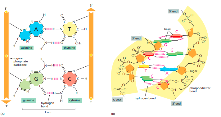
A molecule of deoxyribonucleic acid (DNA) consists of two long polynucleotide chains. Each chain, or strand, is composed of four types of nucleotide subunits, and the two strands are held together by hydrogen bonds between the base portions of the nucleotides. For the nucleotides in DNA, the sugar is deoxyribose (hence the name deoxyribonucleic acid), and the base can be either adenine (A), cytosine (C), guanine (G), or thymine (T). The nucleotides are covalently linked together in a chain through the sugars and phosphates, which thus form a backbone of alternating sugar–phosphate–sugar–phosphate.
BONDS IN DNA
The two strands of the DNA double helix are held together by hydrogen bonds between complementary base pairs. (A) The shapes and chemical structure of the bases allow hydrogen bonds to form efficiently only between A and T and between G and C, where atoms that are able to form hydrogen bonds can be brought close together without perturbing the double helix. Two hydrogen bonds form between A and T, whereas three form between G and C. The bases can pair in this way only if the two polynucleotide chains that contain them are antiparallel—that is, oriented in opposite directions. The nucleotides are linked together covalently by phosphodiester bonds through the 3’-hydroxyl (–OH) group of one sugar and the 5’-phosphate (–OPO3) of the next. This linkage gives each polynucleotide strand a chemical polarity; that is, its two ends are chemically different. The 3’ end carries an unlinked –OH group attached to the 3’ position on the sugar ring; the 5’ end carries a free phosphate group attached to the 5’ position on the sugar ring.
Another important clue to the structure of DNA came from the work of Erwin Chargaff and his colleagues in the late 1940s. They found that the four nucleotide bases of DNA occur in different ratios in the DNAs of different organisms and that the amounts of certain bases are closely related. These data, collected from DNAs of a great many different species, led Chargaff to the following conclusions:
1. The base composition of DNA generally varies from one species to another.
2. DNA specimens isolated from different tissues of the same species have the same base composition.
3. The base composition of DNA in a given species does not change with an organism’s age, nutritional state, or changing environment.
4. In all cellular DNAs, regardless of the species, the number of adenosine residues is equal to the number of thymidine residues (that is, A = T), and the number of guanosine residues is equal to the number of cytidine residues (G = C). From these relationships it follows that the sum of the purine residues equals the sum of the pyrimidine residues; that is, A + G = T + C.
These quantitative relationships, sometimes called “Chargaff’s rules,”
PHYSICAL PROPERTIES ASSOCIATED WITH DNA STRUCTURE
The bases are nearly perpendicular to the helix axis, and adjacent bases are separated by 3.4 Å. The helical structure repeats every 34 Å, so there are 10 bases ( 34 Å per repeat/3.4 Å per base) per turn of helix. There is a rotation of 36 degrees per base (360 degrees per full turn/10 bases per turn). 4. The diameter of the helix is 20 Å.
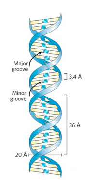
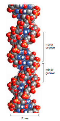
The positions of the base pair relative to the helix axis are described by another three parameter:
BASE PAIR TILT
It is a shift of base pair short axis relative to the vertical helix axis. The tilt angle is measured by considering the angle made by a line drawn through two hydrogen bonded bases relative to a line drawn perpendicular to the helix axis. The tilt angle opens in a direction of the phosphate backbone.
BASE PAIR ROLL
It is a shift of the base pair long axis relative to the vertical helix axis.
PROPELLER TWIST
It is the twist of the bases in a base pair against each other. A base pair is rarely a perfect flat plane with each base in a same plane. Rather, each base has a slightly different roll angle with respect to other base.
TYPES OF DNA DOUBLE HELIX
B FORM OF DNA DOUBLE HELIX
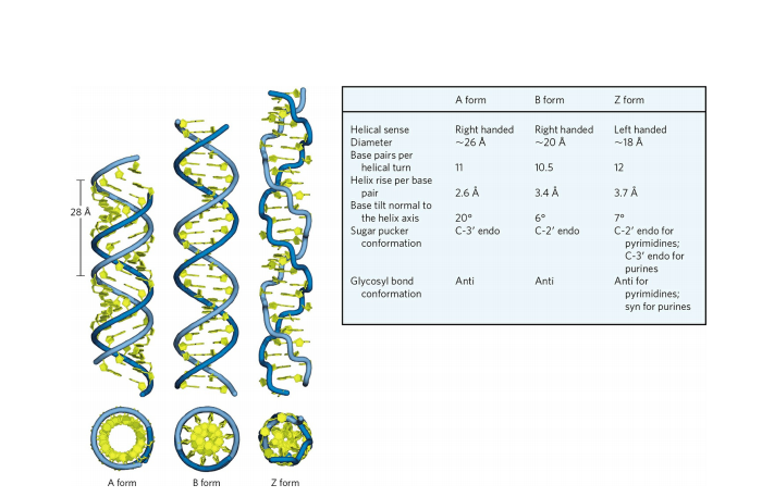
The Watson-Crick structure is also referred to as B-form DNA, or BDNA. The B form is the most stable structure for a random-sequence DNA molecule under physiological conditions and is therefore the standard point of reference in any study of the properties of DNA. Two structural variants that have been well characterized in crystal structures are the A and Z forms.
A FORM OF DNA DOUBLE HELIX
The A form is favored in many solutions that are relatively devoid of water. The DNA is still arranged in a right-handed double helix, but the helix is wider and the number of base pairs per helical turn is 11, rather than 10.5 as in B-DNA. The plane of the base pairs in A-DNA is tilted about 20° relative to B-DNA base pairs, thus the base pairs in A-DNA are not perfectly perpendicular to the helix axis. These structural changes deepen the major groove while making the minor groove shallower. The reagents used to promote crystallization of DNA tend to dehydrate it, and thus most short DNA molecules tend to crystallize in the A form
Z FORM OF DNA DOUBLE HELIX
Z-form DNA is a more radical departure from the B structure; the most obvious distinction is the left-handed helical rotation. There are 12 base pairs per helical turn, and the structure appears more slender and elongated. The DNA backbone takes on a zigzag appearance. Certain nucleotide sequences fold into left-handed Z helices much more readily than others. Prominent examples are sequences in which pyrimidines alternate with purines, especially alternating C and G (that is, in the helix, alternating C≡G and G≡C pairs) or 5-methyl-C and G residues. To form the left-handed helix in ZDNA, the purine residues flip to the syn conformation, alternating with pyrimidines in the anti conformation. The major groove is barely apparent in Z-DNA, and the minor groove is narrow and deep.
CONLUSION
The genes of every cell on Earth are made of DNA, and insights into the relationship between DNA and genes have come from experiments in a wide variety of organisms. Genes and other important segments of DNA are arranged in the single, long DNA molecule that forms the core of each chromosome in the cell
We now know that the DNA carries the hereditary information of the cell and that the protein components of chromosomes function largely to package and control the enormously long DNA molecules. But biologists in the 1940s had difficulty accepting DNA as the genetic material because of the apparent simplicity of its chemistry. DNA, after all, is simply a long polymer composed of only four types of nucleotide subunits, which are chemically very similar to one another. Then, early in the 1950s, DNA was examined by X-ray diffraction analysis, a technique for determining the three-dimensional atomic structure of a molecule. The early results indicated that DNA is composed of two strands wound into a helix. The observation that DNA is double-stranded was of crucial significance. It provided one of the major clues that led, in 1953, to a correct model for the structure of DNA. This structure immediately suggested how DNA could encode the instructions necessary for life, and how these instructions could be copied and passed along when cells divide.
REFERENCE
IMAGES AND CONTENT ARE TAKEN FROM
(A) Lehninger principles of biochemistry seventh edition By David L. Nelson and Michael M. Cox
See Other Posts Also
- STRUCTURE OF AMINO ACIDS AND PEPTIDE
- STRUCTURE AND TYPES OF NUCLEOTIDES
- STRUCTURE OF PROTEIN – PRIMARY, SECONDARY, TERTIARY AND QUATERNARY
- Hantavirus: Symptoms, Causes, Prevention
- NANOPARTICLES
Discover more from Zoologyverse
Subscribe to get the latest posts sent to your email.