
Histology of Testis and Ovary of Lepus
The histology of testis and ovary of Lepus is one of the most common topics in zoology. You meet it in practical exams, viva questions and theory papers. Many students find it hard at first. The slides are full of tiny cells, odd names and similar-looking layers. It is easy to mix them up.
But the topic follows a clear pattern. The testis is built for making sperm all the time. The ovary is built for growing eggs in stages. Once you see this, each layer and each cell has a clear job. The names start to make sense, and the slides stop looking like a puzzle.
Lepus is the scientific name for the hare. Hares belong to the order Lagomorpha, the same group as rabbits. They are mammals, so their gonads show the typical mammalian plan. That makes them a good model for learning how reproductive organs are built. A quick note: many colleges use the rabbit (Oryctolagus) in place of the hare in practicals. The gonads of both animals look almost the same, so these notes work for either one.
In this guide to the histology of testis and ovary of Lepus, you will learn:
- The coverings and lobules of the testis
- The cells of the seminiferous tubule, including Sertoli and Leydig cells
- The layers of the ovary, from germinal epithelium to medulla
- The stages of follicle growth, from primordial follicle to corpus luteum
- A side-by-side comparison of the two organs
- Easy tips to identify each slide and draw neat diagrams
Why Study the Histology of Testis and Ovary of Lepus?
Gonads are the organs that make gametes and sex hormones. In males, the gonad is the testis. In females, it is the ovary. Each one has two big jobs. It must produce sex cells, and it must release hormones that control the reproductive system.
Histology is the study of tissues under a microscope. It shows how an organ’s structure matches its function. When you look at a slide of the testis, you can see the exact spot where sperm are made. When you look at an ovary, you can see eggs at different stages of growth. Nothing is left to guesswork.
There are four main reasons this topic matters.
1. It is a core part of zoology courses.
Students of B.Sc., M.Sc. and entrance exams meet it again and again. Questions can ask for a labelled diagram, a short note, or a comparison between the two organs. Knowing the histology of testis and ovary of Lepus gives you easy marks in all three formats.
2. It explains how mammals reproduce.
Sperm formation, egg growth, ovulation and hormone control all happen inside these tissues. If you understand the slides, you understand the process. Topics like spermatogenesis, oogenesis and the menstrual or oestrous cycle become much simpler.
3. It builds slide-identification skills.
Practical exams often show a slide and ask you to identify it in a minute or two. You need to spot the key features fast. Round tubules mean testis. A fluid-filled follicle means ovary. With practice, this takes seconds.
4. It links to real-life biology and medicine.
Problems like low sperm count, ovarian cysts and hormone imbalance all begin at the tissue level. Doctors and researchers study gonad histology to understand them. Even a basic grasp of the normal structure helps you see what can go wrong.
Why Lepus Is a Good Example
Lepus is a mammal with a typical mammalian reproductive system. Its testis shows clear seminiferous tubules with every stage of sperm development. Its ovary shows follicles from small to mature, plus the corpus luteum. This means one slide can teach you many features at once.
Hares and rabbits are also induced ovulators. Mating triggers the release of the egg, so the ovary can show interesting changes after mating. This makes the topic a little more fun than a plain textbook diagram.
Histology of Testis of Lepus
The testes of a hare are paired, oval organs. They hang in the scrotal sacs, outside the main body cavity. This matters because sperm need a temperature slightly lower than body heat. In hares and rabbits the inguinal canal stays open. So the testes can move up into the abdomen outside the breeding season and drop back down when breeding begins. also see Structure of Male Reproductive System of Lepus
A transverse section (T.S. of testis hare) shows a firm capsule, many small compartments and a crowd of round tubules. Let us go through each part.
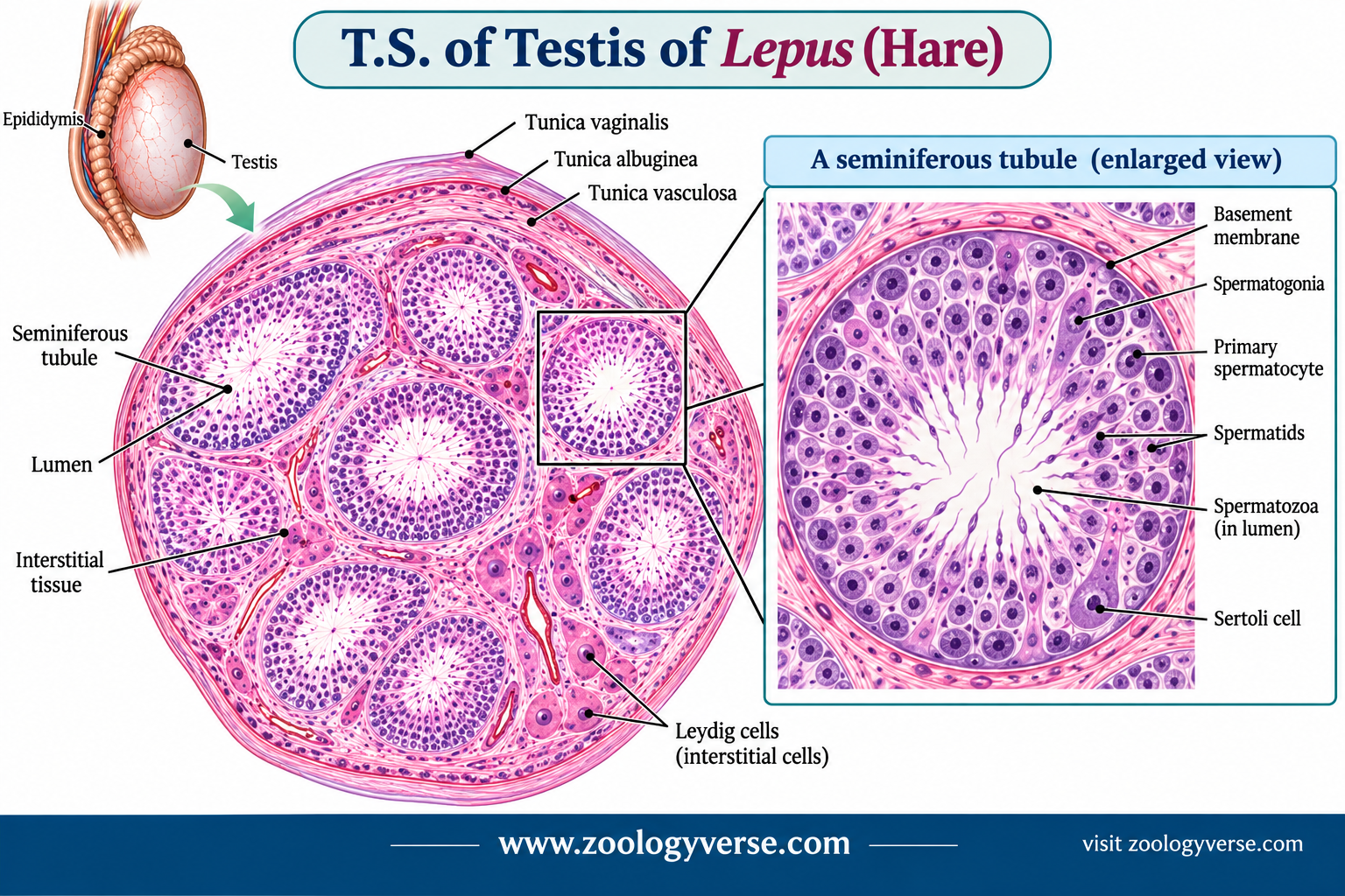
Outer coverings
- Tunica vaginalis: a thin outer layer that comes from the peritoneum. It lets the testis move smoothly.
- Tunica albuginea: a tough white capsule of dense connective tissue. It gives the testis its shape and strength.
- Tunica vasculosa: a soft inner layer packed with blood capillaries. It feeds the tissue below.
Mediastinum and lobules
At the back of the testis, the tunica albuginea thickens into the mediastinum testis. Thin walls called septula testis run inward from it like the spokes of a wheel. They divide the testis into many small lobules. Each lobule holds one to a few tightly coiled seminiferous tubules. If you stretched them out, the tubules would be very long. Coiling lets many of them fit in a small space.
Seminiferous tubules: the sperm factory
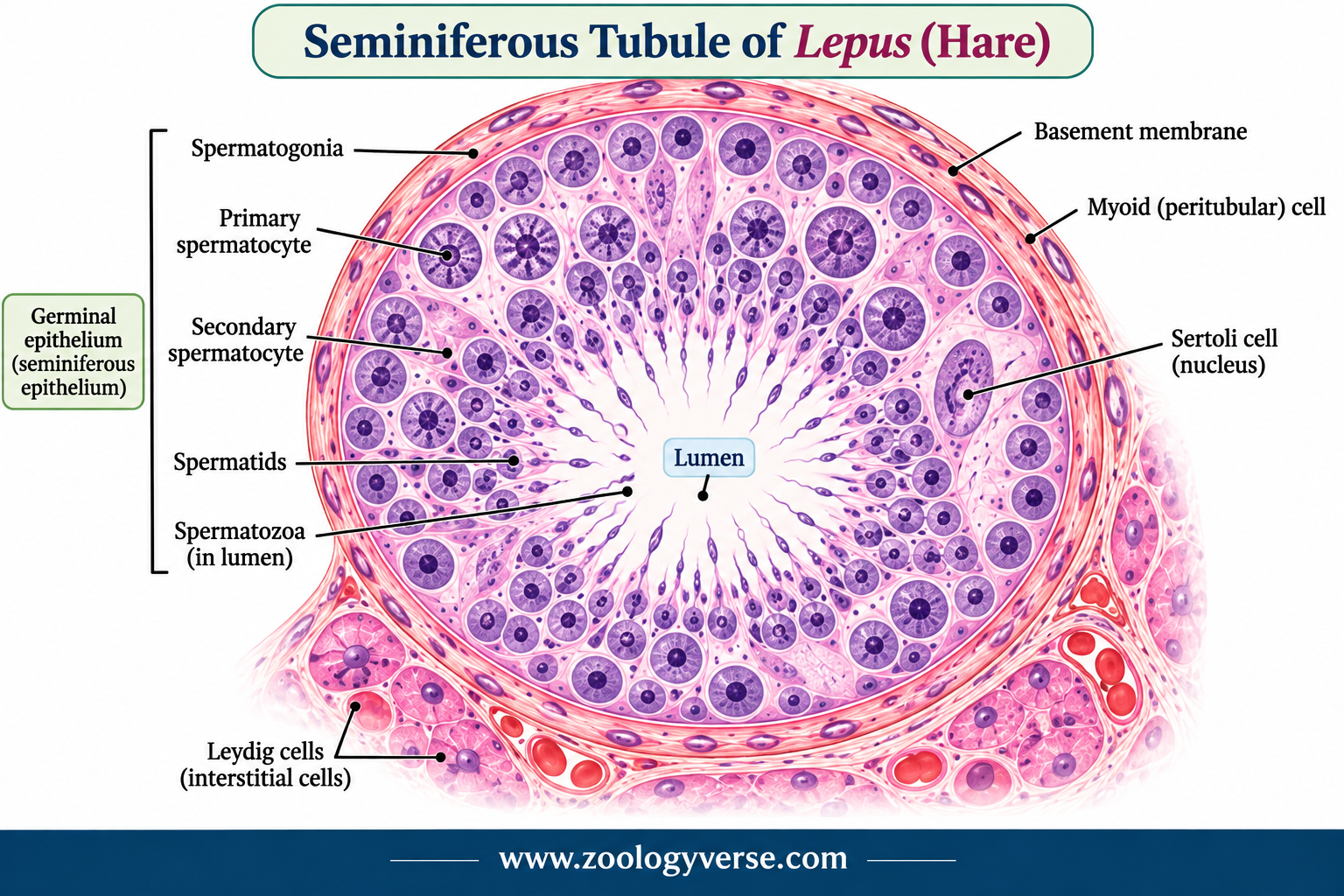
In a section, the tubules look like round or oval rings. Each ring has a hollow centre called the lumen. The wall has three parts:
- A thin basement membrane.
- Flat myoid (peritubular) cells around the outside. Their gentle squeezing helps push sperm along.
- The germinal epithelium, which holds the working cells.
Spermatogenic cells
These cells form layers. The youngest sit at the outer edge. The oldest sit near the lumen. So the layers show the whole story of sperm formation in one picture.
- Spermatogonia: the stem cells. They lie against the basement membrane and divide by mitosis. Some stay as stem cells. Others move on to become sperm.
- Primary spermatocytes: large cells with thick, visible chromosomes. They start meiosis I.
- Secondary spermatocytes: small cells that finish meiosis II very quickly. This is why they are rare on a slide.
- Spermatids: small, round cells with a single set of chromosomes. They do not divide again. They change shape instead.
- Spermatozoa: the mature sperm. The head holds the nucleus and a cap called the acrosome. The tail points into the lumen.
The change from spermatid to sperm is called spermiogenesis. The cell loses most of its cytoplasm, grows a tail and builds the acrosome. The leftover cytoplasm is cleared away by Sertoli cells.
Sertoli cells: the support team
Sertoli cells are tall and irregular. They stretch from the basement membrane to the lumen. Their nuclei are pale, with a clear nucleolus, and usually sit near the base. You can spot them because they do not look like the crowded, dark germ cells around them.
Their jobs are many:
- They feed and support the developing sperm.
- They form tight junctions with neighbouring Sertoli cells. This builds the blood-testis barrier. It splits the tubule into an outer and an inner zone and protects the developing sperm from the immune system.
- They eat the leftover cytoplasm shed during spermiogenesis.
- They help release mature sperm into the lumen.
- They make inhibin and androgen-binding protein. FSH controls them.
Leydig cells: the hormone makers
The space between tubules is called the interstitial tissue. It holds loose connective tissue, blood vessels, lymph vessels and nerves. It also holds clusters of Leydig cells. These are large, polygonal cells with a round nucleus and pale, foamy cytoplasm. They stand out as pink groups between the tubules.
LH tells them to make testosterone. This hormone keeps sperm production going. It also shapes male body features and behaviour.
The path out of the testis
Mature sperm leave the seminiferous tubules and enter the tubuli recti. From there they pass through the rete testis, then the efferent ductules, and finally the epididymis. Sperm mature and are stored there until needed.ubules through the tubuli recti. They then pass through the rete testis and efferent ductules. They finish in the epididymis, where they mature and are stored.
Histology of Ovary of Lepus
The ovary of a hare is a small, oval, solid organ. It is held in place by a fold called the mesovarium. Unlike the testis, the ovary changes a lot over the cycle. One section can show tiny follicles, large follicles and old scars all at once. This makes the histology of ovary of Lepus both rich and a little tricky. also see Structure of Female Reproductive System of Lepus.
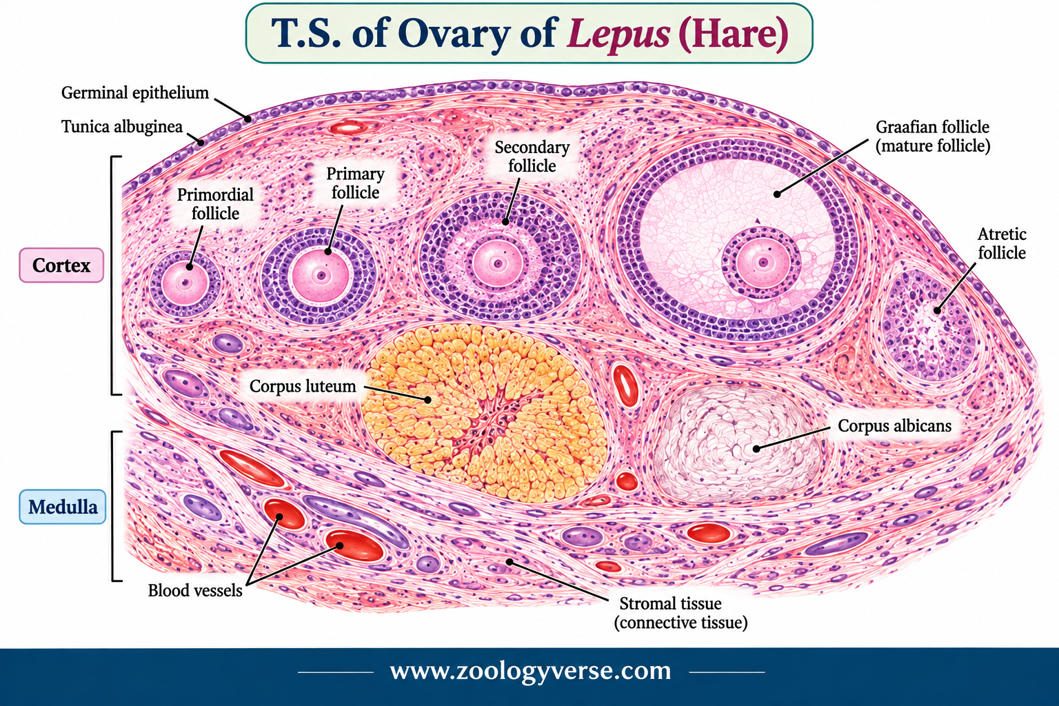
A T.S. of ovary hare shows these layers from outside to inside.
Germinal epithelium
The outermost layer is a single row of cube-shaped cells. It is a modified part of the peritoneum. The name is misleading. It does not produce eggs. Early anatomists gave it that name by mistake, and it stuck.
Tunica albuginea
Just below the epithelium is a thin band of dense connective tissue. It protects the ovary and separates the surface from the cortex.
Cortex: where the follicles live
The cortex is the thick outer zone. It is made of stroma, a cell-rich connective tissue with spindle-shaped cells. Follicles at all stages lie inside it. A follicle is an egg cell plus the cells that surround and feed it.
Primordial follicle. The smallest and most common type. It has a primary oocyte wrapped in one layer of flat follicle cells. The oocyte is paused in prophase of meiosis I. It stays paused, sometimes for a long time, until the follicle is called to grow.
Primary follicle. The oocyte grows bigger. The flat cells become cube-shaped and then pile into layers. A clear, jelly-like coat called the zona pellucida forms around the oocyte.
Secondary follicle. The layers of granulosa cells thicken. Small fluid-filled spaces start to appear. The stroma around the follicle splits into two layers:
- Theca interna: cells rich in blood vessels. They make hormone precursors under LH.
- Theca externa: a tougher outer layer of fibres that holds the follicle in place.
Graafian (tertiary) follicle. The mature follicle, and the star of the slide. The small spaces merge into one large cavity, the antrum, filled with liquor folliculi. The oocyte sits on a small mound of cells, the cumulus oophorus. A ring of cells right around the oocyte forms the corona radiata. The granulosa cells turn the hormone precursors into oestrogen. FSH drives this step.
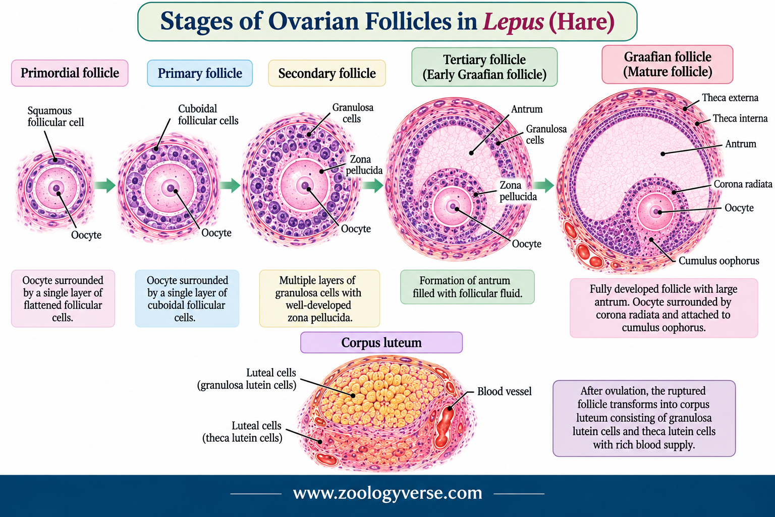
Just before ovulation, the primary oocyte finishes meiosis I and becomes a secondary oocyte. It then pauses again until a sperm enters.
Corpus luteum. After the egg is released, the burst follicle collapses and turns into a yellowish gland. Its large cells make progesterone. If the egg is not fertilised, rabbits and hares can go through a pseudopregnancy before the gland fades.
Corpus albicans. When the corpus luteum fades, it shrinks to a pale scar of connective tissue.
Atretic follicles. Most follicles never ovulate. They break down at some stage. This is called atresia, and you will see it on almost every slide.
Induced ovulation
Hares and rabbits are usually described as induced ovulators. The egg is not released on a fixed schedule. Mating triggers the release, some hours later. So the follicles stay ready, and corpora lutea appear mainly after mating. This is a useful point for exam answers.
Medulla
The inner zone is loose connective tissue. It holds large, coiled blood vessels, nerves and lymph vessels. There are no follicles here. Its main job is to supply the cortex.
Testis vs Ovary of Lepus: Quick Comparison
| Feature | Testis | Ovary |
|---|---|---|
| Outer covering | Tunica vaginalis and tunica albuginea | Germinal epithelium and tunica albuginea |
| Functional units | Seminiferous tubules | Ovarian follicles |
| Gametes produced | Spermatozoa (continuous) | Ova (cyclical) |
| Supporting cells | Sertoli cells | Follicle (granulosa) cells |
| Hormone-secreting cells | Leydig cells (testosterone) | Theca and granulosa cells (oestrogen), corpus luteum (progesterone) |
| Zones | Lobules and interstitial tissue | Cortex and medulla |
How to Identify the Slides Quickly
Spotting the right slide is half the marks in a practical. For the histology of testis and ovary of Lepus, use these clues:
- Round tubules with layered cells and an open centre: testis.
- A big fluid-filled cavity with an egg on a small stalk: Graafian follicle, so ovary.
- Small pink cell clusters between tubules: Leydig cells.
- A yellow, folded mass of large cells: corpus luteum.
Drawing Tips for Exams
Keep your diagram neat and large. Use a sharp pencil and straight label lines. For the testis, label the tunica albuginea, seminiferous tubule, Sertoli cell, spermatogonia, spermatozoa and Leydig cells. For the ovary, label the germinal epithelium, primordial follicle, Graafian follicle, oocyte, antrum, corpus luteum and medulla.
Key Takeaways
- The testis of Lepus has lobules of coiled seminiferous tubules.
- Sertoli cells support sperm and form the blood-testis barrier.
- Leydig cells make testosterone.
- The ovary has a follicle-rich cortex and a vascular medulla.
- The Graafian follicle is the main landmark of a mature ovary.
- Learning the histology of testis and ovary of Lepus is mostly about knowing these cells and layers.
Frequently Asked Questions
What do Sertoli cells do in the histology of testis and ovary of Lepus?
They are found in the testis. They feed sperm cells, protect them and form the blood-testis barrier.
Where are Leydig cells found?
In the connective tissue between the seminiferous tubules. They secrete testosterone.
What is the difference between a primary follicle and a Graafian follicle?
A primary follicle is small, with a growing oocyte and a single layer of cuboidal cells. A Graafian follicle is large and mature, with a fluid-filled antrum and a fully developed oocyte ready for ovulation.
Why is the ovary’s outer layer called germinal epithelium?
It is an old name based on the mistaken belief that it produced eggs. It is really a simple cuboidal covering layer.
What is the corpus luteum?
A temporary endocrine structure formed from the ruptured follicle after ovulation. It secretes progesterone.
Final Thoughts on the Histology of Testis and Ovary of Lepus
The testis shows continuous sperm production inside tubules. The ovary shows eggs growing in follicles, stage by stage. Learn the layers, learn the key cells, and practise your labelled diagrams. Then any slide in this topic will feel familiar.
References and Further Reading
- Junqueira, L. C. and Carneiro, J., Basic Histology: Text and Atlas
- Ross, M. H. and Pawlina, W., Histology: A Text and Atlas
- Kardong, K. V., Vertebrates: Comparative Anatomy, Function, Evolution
- Jordan, E. L. and Verma, P. S., Chordate Zoology
Discover more from Zoologyverse
Subscribe to get the latest posts sent to your email.