
The Complement System is an important part of the immune system that helps the body recognize and eliminate invading microorganisms. It consists of a group of soluble proteins and cell-surface proteins present in blood plasma and other body fluids. These proteins work together in a coordinated cascade to strengthen immune defence against bacteria, viruses, fungi, and other potentially harmful agents.
The Complement System is closely associated with innate immunity, which provides rapid, non-specific protection against infections. It also supports adaptive immunity by enhancing antibody-mediated immune responses. When activated, complement proteins can mark pathogens for destruction, promote inflammation, attract immune cells to infected tissues, and directly damage certain microorganisms by forming the membrane attack complex (MAC).
The system can be activated through three major pathways: the classical pathway, the alternative pathway, and the lectin pathway. Although these pathways begin in different ways, they converge on the activation of complement component C3 and contribute to a common set of immune responses.
The Complement System is essential for studying immunology because it connects pathogen recognition, inflammation, phagocytosis, and direct microbial killing. Its activity must also be carefully regulated to prevent unnecessary damage to the body’s own cells.
Definition of the Complement System
The Complement System is a group of plasma proteins and membrane-associated proteins that interact in a regulated cascade to enhance the body’s immune defence against pathogens and abnormal cells. Most complement proteins circulate in the blood in inactive forms and become activated in a specific sequence when triggered by microorganisms, antibodies, or other immune-related signals.
The term complement refers to the ability of these proteins to complement or strengthen the actions of antibodies and immune cells. The system is a major component of innate immunity, but it also contributes to adaptive immunity by supporting antibody-mediated responses.
Once activated, the Complement System produces several important effects, including:
- Opsonization: Coating pathogens with complement fragments, particularly C3b, to make them easier for phagocytes to recognize and engulf.
- Inflammation: Releasing complement fragments such as C3a and C5a, which promote inflammatory responses.
- Chemotaxis: Attracting immune cells, especially through the action of C5a, toward sites of infection.
- Cell lysis: Forming the membrane attack complex (MAC), which can create pores in susceptible microbial membranes.
- Immune-complex clearance: Helping remove antigen–antibody complexes and certain cellular debris from circulation.
Complement activation follows a cascade mechanism in which one activated component helps activate the next. This amplifies the immune response and enables the body to respond efficiently to potential threats. However, regulatory proteins control this process to limit damage to healthy host tissues.
Components of the Complement System
The Complement System consists of a large group of plasma proteins, membrane-bound proteins, and regulatory molecules that work together to protect the body against infections. Most complement proteins are produced by the liver, although some immune cells and other tissues also contribute to their production. These components circulate in inactive forms and become activated when the immune system detects an appropriate trigger.
The major components can be grouped according to their roles in complement activation, amplification, pathogen destruction, and regulation.
1. Complement Components C1–C9
The classical complement pathway involves components C1, C2, C3, C4, and C5, while the terminal stages involve C5, C6, C7, C8, and C9. C3 is particularly important because its activation generates fragments that promote opsonization and inflammation and help initiate the terminal pathway.
| Component | Major function |
|---|---|
| C1 | Initiates the classical pathway through recognition of antibody-bound targets. |
| C2 | Participates in forming the classical and lectin pathway C3 convertase. |
| C3 | Produces C3a and C3b; supports inflammation, opsonization, and pathway progression. |
| C4 | Participates in classical and lectin pathway activation. |
| C5 | Produces C5a, a potent inflammatory mediator, and C5b, which initiates MAC formation. |
| C6 | Joins the terminal complex during MAC assembly. |
| C7 | Helps anchor the developing terminal complex to the target membrane. |
| C8 | Contributes to membrane insertion and pore formation. |
| C9 | Polymerizes to help form the pore of the membrane attack complex. |
2. Factors of the Alternative Pathway
The alternative pathway uses additional proteins that help activate and amplify complement responses on suitable microbial surfaces.
- Factor B: Binds to C3b and participates in forming the alternative pathway C3 convertase after activation by Factor D.
- Factor D: Cleaves Factor B when it is associated with C3b.
- Properdin (Factor P): Stabilizes the alternative pathway C3 convertase and helps sustain complement activation.
These factors enable the alternative pathway to amplify complement activity without requiring antibodies to initiate the response.
3. Lectin Pathway Recognition Proteins
The lectin pathway recognizes particular carbohydrate patterns on microbial surfaces.
- Mannose-binding lectin (MBL): Binds to certain sugar patterns found on microorganisms.
- Ficolins: Recognize specific molecular patterns on microbial surfaces.
- MASPs (MBL-associated serine proteases): Become activated following recognition and help cleave C4 and C2, initiating the downstream complement cascade.
The lectin pathway therefore provides a way to recognize pathogens without requiring antibodies.
4. Regulatory Proteins
The Complement System must be carefully controlled because excessive activation can damage healthy tissues. Regulatory proteins limit activation, accelerate the breakdown of convertases, or prevent inappropriate membrane attack complex formation.
Important regulators include:
- C1 inhibitor (C1-INH): Controls activation of C1 and also regulates components of the lectin pathway.
- Factor H: Helps regulate the alternative pathway, particularly on host surfaces.
- Factor I: Inactivates C3b and C4b with the help of appropriate cofactors.
- DAF (CD55): Accelerates the decay of complement convertases.
- CD59: Inhibits the final stages of membrane attack complex formation on host cells.
These regulatory mechanisms help distinguish the body’s own cells from invading microorganisms and reduce the risk of complement-mediated tissue injury.
The complement proteins act as an interconnected defence network. Recognition proteins initiate activation, enzymatic components amplify the response, terminal components can damage susceptible pathogens, and regulatory proteins protect host tissues. Together, they enable the Complement System to support innate and adaptive immune responses.
Activation of the Complement System
The Complement System is activated through a series of controlled biochemical reactions known as a complement cascade. In this process, the activation of one protein triggers the activation of other components, producing an amplified immune response. This mechanism enables the body to recognize pathogens, promote inflammation, and eliminate susceptible microorganisms efficiently.
Complement proteins normally circulate in blood plasma in inactive forms. When an appropriate trigger is detected, specific proteins are cleaved or undergo structural changes, allowing the next steps of the cascade to occur.
Three Pathways of Complement Activation
The Complement System can be activated through three major pathways:
- Classical pathway: Usually initiated when C1q recognizes antibodies, particularly IgM or certain IgG subclasses, bound to their target antigens. It can also be activated by some antibody-independent targets.
- Alternative pathway: Initiated through spontaneous low-level C3 activation and amplified on suitable microbial surfaces. It does not require antibodies for activation.
- Lectin pathway: Initiated when mannose-binding lectin or ficolins recognize particular molecular patterns on microorganisms. Associated proteases then activate downstream complement components.
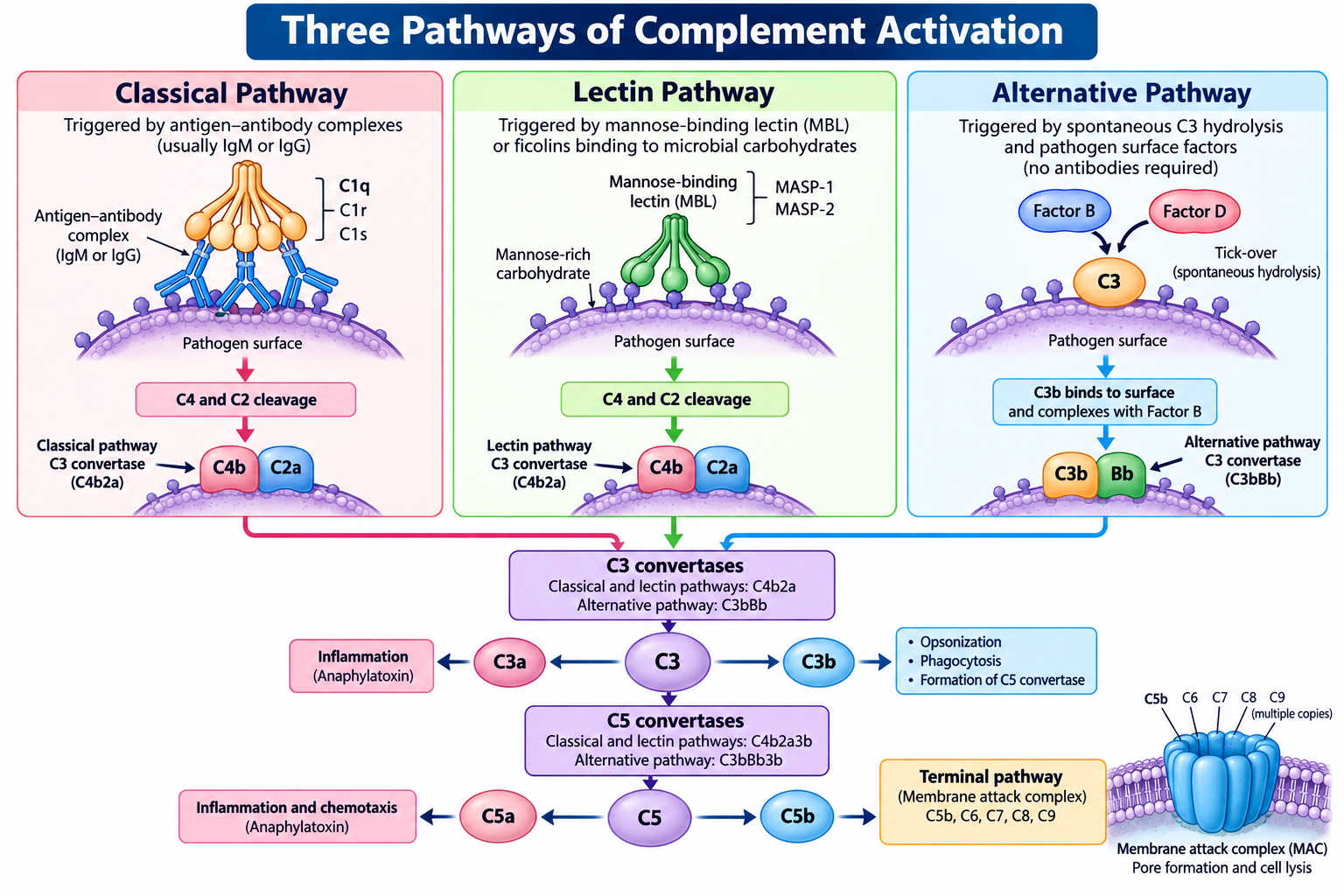
Although these pathways have different initiation mechanisms, the classical and lectin pathways converge through the formation of a C3 convertase containing C4b and C2a. The alternative pathway forms a different C3 convertase containing C3b and Bb.
Common Steps in Complement Activation
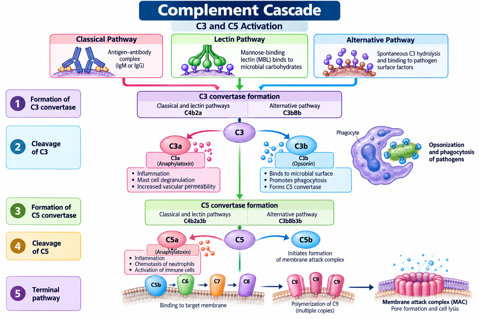
The three pathways eventually contribute to a common sequence of immune reactions.
Step 1: Formation of C3 convertase
Each pathway generates an enzyme complex called C3 convertase. This enzyme cleaves complement component C3 into C3a and C3b.
Step 2: Opsonization and inflammation
C3b attaches to suitable surfaces and acts as an opsonin, helping phagocytes recognize and engulf pathogens. C3a contributes to inflammatory responses.
Step 3: Formation of C5 convertase
Additional C3b associates with the pathway-specific convertase to form C5 convertase. This enzyme cleaves C5 into C5a and C5b.
Step 4: Initiation of the terminal pathway
C5b recruits C6, C7, C8, and C9 to assemble the membrane attack complex (MAC). This complex forms a pore in susceptible target membranes and can cause cell lysis.
Regulation During Activation
Complement activation must remain controlled. Regulatory proteins limit excessive enzyme activity, inactivate selected complement fragments, and protect host cell membranes from inappropriate damage. Without effective regulation, complement activation may contribute to inflammation and tissue injury.
The three pathways differ in how they begin, but their shared downstream reactions allow complement activation to coordinate pathogen recognition, inflammation, opsonization, and direct microbial killing.
Classical Pathway of Complement Activation
The classical pathway is one of the three major routes through which the Complement System becomes activated. It is commonly associated with adaptive immunity because it is often initiated by antibodies bound to antigens. However, certain antibody-independent interactions can also activate this pathway.
The classical pathway helps connect antibody-mediated recognition with complement-mediated immune defence. Once activated, it promotes opsonization, inflammation, and the elimination of susceptible pathogens.
Initiation of the Classical Pathway
The pathway usually begins when the C1 complex recognizes antibodies attached to their target antigens. The C1 complex consists of C1q, C1r, and C1s.
- C1q: Recognizes the Fc regions of appropriately arranged antibodies bound to antigens.
- C1r: Becomes activated following recognition by C1q and activates C1s.
- C1s: Cleaves complement components C4 and C2, allowing the pathway to progress.
IgM is particularly effective at activating the classical pathway when bound to an antigen. Certain IgG subclasses can also activate it, depending on their arrangement and the species involved.
Formation of C3 Convertase
After activation, C1s cleaves C4 into C4a and C4b. C4b attaches to a nearby surface, and C2 binds to it before being cleaved by C1s.
The resulting C4b2a complex acts as the C3 convertase of the classical pathway. It cleaves C3 into two fragments:
- C3a: Contributes to inflammatory responses.
- C3b: Binds to suitable surfaces, promotes opsonization, and helps advance complement activation.
Formation of C5 Convertase and the Terminal Pathway
When additional C3b associates with the C3 convertase, the complex becomes the classical pathway C5 convertase, commonly designated C4b2a3b.
This enzyme cleaves C5 into C5a and C5b. C5a is a potent inflammatory mediator and chemoattractant, whereas C5b initiates the assembly of the membrane attack complex (MAC) by recruiting C6, C7, C8, and C9.
Functions of the Classical Pathway
The classical pathway contributes to immune defence in several ways:
- Antibody-assisted recognition: Connects antigen-bound antibodies with complement activation.
- Opsonization: C3b and its breakdown products help phagocytes recognize and remove pathogens.
- Inflammation: C3a and C5a contribute to inflammatory responses.
- Target-cell damage: Terminal complement components can form the MAC on susceptible membranes.
- Immune-complex clearance: Complement fragments help facilitate the removal of antigen–antibody complexes.
The classical pathway therefore plays an important role in coordinating antibody recognition with the effector functions of the Complement System. Its activity is regulated by several inhibitory proteins to limit damage to healthy tissues.
Alternative Pathway of Complement Activation
The alternative pathway is one of the three major pathways of the Complement System. Unlike the classical pathway, it does not require antibodies to initiate activation. Instead, it depends on the continuous, low-level spontaneous activation of complement component C3 and the amplification of complement activity on suitable microbial surfaces.
This pathway is an important part of innate immunity because it can respond rapidly to invading microorganisms. It also amplifies complement activation initiated through the classical and lectin pathways.
Initiation of the Alternative Pathway
A small amount of C3 undergoes spontaneous hydrolysis in plasma, producing a form that can participate in the alternative pathway. This process is commonly called tick-over. Through subsequent reactions, C3b can be generated and deposited on nearby surfaces.
When C3b binds to a suitable microbial surface, it can associate with Factor B. Factor D then cleaves Factor B into Ba and Bb. The Bb fragment remains associated with C3b, forming the alternative pathway C3 convertase, C3bBb.
Formation of C3 and C5 Convertases
The C3 convertase cleaves additional C3 molecules into C3a and C3b. C3a contributes to inflammation, while C3b supports opsonization and increases complement activation by helping generate more C3 convertase.
The protein properdin (Factor P) stabilizes the C3bBb complex, allowing it to remain active for longer.
When an additional C3b molecule associates with the C3 convertase, the resulting complex acts as the alternative pathway C5 convertase, commonly written as C3bBbC3b.
This enzyme cleaves C5 into:
- C5a: A potent inflammatory mediator that attracts and activates immune cells.
- C5b: Initiates the terminal complement sequence leading to membrane attack complex formation.
Major Components Involved
| Component | Function |
|---|---|
| C3 | Provides C3b for surface deposition and pathway amplification. |
| Factor B | Supplies the Bb fragment of the alternative pathway convertase. |
| Factor D | Cleaves Factor B when it is bound to C3b. |
| Properdin | Stabilizes the C3 convertase. |
| Factor H | Helps regulate alternative pathway activation, especially on host surfaces. |
| Factor I | Inactivates C3b with appropriate cofactors. |
Biological Importance
The alternative pathway contributes to immune defence in several ways:
- Rapid innate defence: It can activate without antibodies.
- Amplification: It increases C3b deposition and strengthens complement responses initiated by any of the three pathways.
- Opsonization: C3b and its breakdown products help phagocytes recognize and engulf pathogens.
- Inflammation: C3a and C5a promote inflammatory responses.
- Microbial killing: The pathway contributes to terminal complement activation and membrane attack complex formation.
Regulation of the Alternative Pathway
The alternative pathway must distinguish microbial surfaces from healthy host tissues. Regulatory proteins such as Factor H and Factor I limit inappropriate C3b activity, while other membrane-associated regulators help protect host cells.
Defects in these regulatory mechanisms can cause excessive complement activation and contribute to inflammatory or tissue-damaging disorders.
The alternative pathway therefore provides an antibody-independent defence mechanism and acts as a powerful amplification system within the Complement System.
Lectin Pathway of Complement Activation
The lectin pathway is one of the three major pathways of the Complement System. It forms an important part of innate immunity because it recognizes characteristic carbohydrate patterns on microbial surfaces without requiring antibodies. This enables the immune system to detect certain pathogens and initiate complement-mediated defence.
The lectin pathway shares several downstream steps with the classical pathway, but its initiation mechanism is different. Instead of relying primarily on antibodies and the C1 complex, it uses carbohydrate-recognition proteins and associated enzymes.
Initiation of the Lectin Pathway
The pathway begins when recognition molecules bind to specific molecular patterns on the surface of microorganisms.
The major recognition molecules include:
- Mannose-binding lectin (MBL): Recognizes certain sugar arrangements, including mannose-rich patterns, on microbial surfaces.
- Ficolins: Recognize particular carbohydrate and acetylated molecular patterns associated with microorganisms.
These recognition molecules are associated with enzymes called MBL-associated serine proteases (MASPs). Binding to an appropriate target activates these proteases and initiates the complement cascade.
Formation of C3 Convertase
Activated MASPs, particularly MASP-2, cleave complement components C4 and C2. The resulting fragments assemble to form the C3 convertase C4b2a, which is also produced by the classical pathway.
This enzyme cleaves C3 into:
- C3a: Contributes to inflammatory responses.
- C3b: Promotes opsonization and supports further complement activation.
The formation of C3 convertase allows the lectin pathway to amplify the immune response against the recognized target.
Formation of C5 Convertase and the Terminal Pathway
Additional C3b associates with the C3 convertase to form the lectin pathway C5 convertase, commonly designated C4b2a3b.
This enzyme cleaves C5 into C5a and C5b. C5a promotes inflammation and attracts immune cells, whereas C5b initiates the assembly of the membrane attack complex (MAC).
The terminal complement components C6, C7, C8, and C9 then assemble with C5b to form a pore in susceptible target membranes.
Functions of the Lectin Pathway
The lectin pathway supports immune defence through several mechanisms:
- Pathogen recognition: Detects characteristic molecular patterns on microbial surfaces.
- Opsonization: C3b and related fragments help phagocytes recognize and engulf pathogens.
- Inflammation: Complement fragments such as C3a and C5a contribute to inflammatory responses.
- Amplification: Activates the shared downstream complement cascade.
- Membrane attack complex formation: Contributes to the destruction of susceptible microorganisms.
Difference Between the Lectin and Classical Pathways
The lectin and classical pathways share the same C3 convertase but differ in how they are initiated.
| Feature | Lectin pathway | Classical pathway |
|---|---|---|
| Main recognition mechanism | Recognition of microbial molecular patterns | Usually recognition of antigen-bound antibodies |
| Important recognition molecules | MBL and ficolins | C1q |
| Associated enzymes | MASPs | C1r and C1s |
| C3 convertase | C4b2a | C4b2a |
| Requirement for antibodies | Not required | Commonly antibody-dependent, but not always |
The lectin pathway is therefore an important antibody-independent mechanism for initiating complement activation. Together with the classical and alternative pathways, it helps the Complement System recognize pathogens and coordinate a protective immune response.
Membrane Attack Complex (MAC)
The membrane attack complex (MAC) is the terminal structure formed during activation of the Complement System. It consists of complement components C5b, C6, C7, C8, and multiple C9 molecules. The complex assembles on a susceptible target membrane and forms a pore that can disrupt membrane integrity and cause cell lysis.
The MAC is particularly important in defence against certain Gram-negative bacteria, including Neisseria species. However, its effectiveness varies with the microorganism and the structure of its outer membrane.
Formation of the Membrane Attack Complex
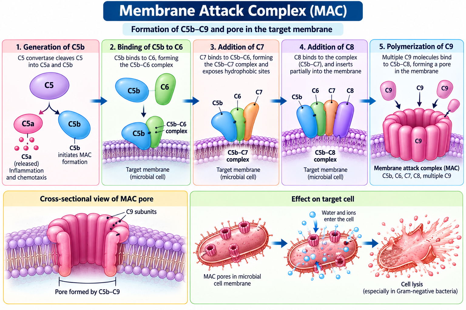
MAC formation begins after C5 convertase cleaves complement component C5 into C5a and C5b. The C5b fragment initiates the assembly of the terminal complement components.
The process occurs in the following sequence:
- Formation of C5b: C5 convertase cleaves C5, generating C5b.
- Binding of C6: C5b binds C6 to form the C5b6 complex.
- Addition of C7: C7 joins the complex, allowing it to associate with the target membrane.
- Binding of C8: C8 associates with the complex and contributes to membrane insertion and pore formation.
- Polymerization of C9: Multiple C9 molecules assemble to form the transmembrane pore.
The resulting C5b-9 complex is known as the membrane attack complex.
Structure of the MAC
The MAC forms a ring-like pore that spans the target membrane. This pore disturbs normal membrane permeability and ion balance. Depending on the target cell and the extent of complement activation, the resulting damage can lead to cell lysis.
Host cells are normally protected from inappropriate MAC formation by regulatory mechanisms. One important regulator is CD59, a membrane-associated protein that inhibits the terminal stages of MAC assembly.
Functions of the Membrane Attack Complex
The major functions of the MAC include:
- Direct microbial killing: Damages susceptible microorganisms by forming membrane pores.
- Membrane disruption: Interferes with normal membrane integrity and ion balance.
- Terminal complement activity: Provides the final effector mechanism shared by all three complement activation pathways.
Clinical Significance
Deficiencies in terminal complement components, particularly C5–C9, can increase susceptibility to recurrent infections caused by Neisseria species. This demonstrates the importance of the MAC in immune protection.
The MAC is a key effector mechanism of the Complement System, but it is not responsible for all complement-mediated pathogen elimination. Opsonization and inflammation also play major roles in immune defence.
Functions of the Complement System
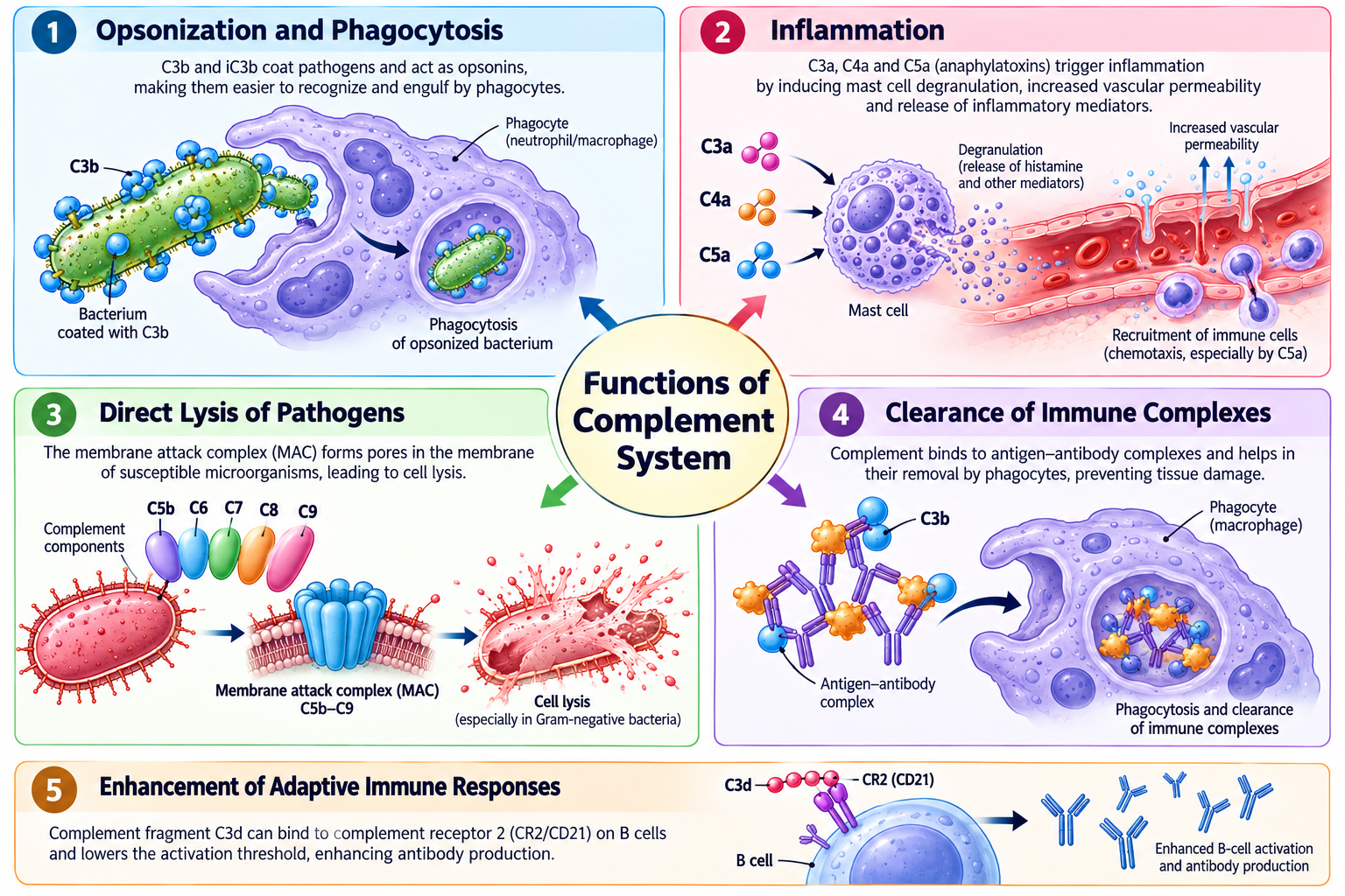
The Complement System performs several important functions that strengthen immune defence and help maintain tissue health. Through a coordinated series of reactions, complement proteins recognize or mark pathogens, promote inflammation, support phagocytosis, and contribute to the removal of immune complexes and damaged cellular material.
The major functions are described below.
1. Opsonization and Phagocytosis
Opsonization is the process of coating a pathogen with molecules that make it easier for phagocytic cells to recognize and engulf it. Complement fragment C3b is an important opsonin, while its breakdown products, including iC3b, also promote recognition by specific complement receptors.
Neutrophils and macrophages recognize these complement-coated targets through complement receptors and engulf them by phagocytosis. This process improves the clearance of many bacteria and other particles.
2. Promotion of Inflammation
Complement activation produces fragments that contribute to inflammatory responses. C3a and C5a are known as anaphylatoxins because they can promote inflammatory processes, including mast-cell activation under appropriate conditions.
These mediators help increase vascular permeability and support the recruitment of immune cells to affected tissues. C5a is also a particularly potent chemoattractant and activator of several leukocytes.
3. Chemotaxis of Immune Cells
Chemotaxis is the directed movement of cells in response to chemical signals. C5a helps attract neutrophils and other immune cells toward sites of infection or tissue injury.
Once recruited, these cells can recognize, engulf, and destroy pathogens, helping coordinate the local immune response.
4. Direct Lysis of Pathogens
The terminal complement components C5b, C6, C7, C8, and C9 assemble to form the membrane attack complex (MAC). The complex creates a pore in susceptible target membranes, which can disrupt membrane integrity and cause cell lysis.
This mechanism is particularly important against certain Gram-negative bacteria, including Neisseria species. Its effectiveness depends on the properties of the target microorganism and its membrane.
5. Clearance of Immune Complexes
Immune complexes are formed when antibodies bind to antigens. If these complexes are not removed efficiently, they may accumulate in tissues and contribute to inflammation.
Complement fragments, particularly C3b, can bind to immune complexes and facilitate their transport and clearance through complement receptors on blood cells and their subsequent removal by phagocytic cells, especially in the liver and spleen.
6. Enhancement of Adaptive Immunity
Although complement is a major component of innate immunity, it also supports adaptive immune responses. The complement fragment C3d can bind to complement receptor CR2 (CD21) on B cells and help enhance B-cell activation when antigen is recognized.
This interaction can lower the threshold for B-cell activation and improve antibody responses under appropriate conditions.
7. Removal of Cellular Debris
Complement proteins can help identify and facilitate the clearance of apoptotic cells and certain forms of cellular debris. Efficient removal of this material helps maintain tissue homeostasis and reduces the likelihood that cellular contents will provoke harmful inflammatory or autoimmune responses.
Summary of Major Functions
| Function | Important complement component or fragment | Main outcome |
|---|---|---|
| Opsonization | C3b, iC3b | Promotes pathogen recognition and phagocytosis |
| Inflammation | C3a, C5a | Supports inflammatory responses |
| Chemotaxis | C5a | Recruits immune cells to affected tissues |
| Direct lysis | C5b–C9 | Forms the membrane attack complex |
| Immune-complex clearance | C3b and related fragments | Facilitates removal of immune complexes |
| Enhancement of B-cell responses | C3d | Supports B-cell activation through CR2 |
| Cellular debris clearance | Complement fragments and receptors | Helps maintain tissue homeostasis |
The Complement System links pathogen recognition with inflammation, phagocytosis, direct microbial killing, and adaptive immune responses. These functions work together to provide effective protection while complement-regulatory mechanisms limit damage to healthy tissues.
Regulation of Complement Activation
The Complement System is a powerful immune defence mechanism, but uncontrolled activation can damage healthy cells and tissues. To prevent this, the body uses several regulatory proteins that control complement activation, inactivate selected complement fragments, and protect host-cell membranes.
Regulation is essential because complement proteins must eliminate pathogens while minimizing unnecessary inflammation and tissue injury.
1. Regulation of the Classical and Lectin Pathways
C1 inhibitor (C1-INH) is an important regulatory protein that controls the activation of the classical and lectin pathways. It inhibits activated C1r and C1s and also regulates the lectin pathway-associated proteases MASP-1 and MASP-2.
By limiting these enzymes, C1-INH helps prevent excessive initiation of complement activation.
2. Regulation of the Alternative Pathway
The alternative pathway requires tight control because it undergoes continuous low-level activation and can amplify complement responses.
Two major regulatory proteins are:
- Factor H: Binds to C3b and helps regulate alternative pathway activation, particularly on host-cell surfaces. It also acts as a cofactor for Factor I.
- Factor I: Cleaves and inactivates C3b and C4b with the help of appropriate cofactors, limiting further complement activation.
These proteins help prevent excessive complement activity and protect healthy tissues.
3. Regulation of C3 and C5 Convertases
Convertases are enzyme complexes that drive complement activation. Several regulatory proteins control their formation or accelerate their breakdown.
- Decay-accelerating factor (DAF or CD55): Promotes the breakdown of C3 and C5 convertases on cell surfaces.
- Complement receptor 1 (CR1 or CD35): Helps regulate convertases and acts as a cofactor for Factor I-mediated cleavage of C3b and C4b.
These mechanisms restrict the duration and intensity of complement activation.
4. Protection Against Membrane Attack Complex Formation
The terminal complement pathway can damage membranes by assembling the membrane attack complex (MAC). Host cells possess protective mechanisms that reduce this risk.
CD59, also known as protectin, inhibits the terminal stages of MAC assembly by interfering with C9 incorporation and polymerization. This reduces the formation of damaging pores in host-cell membranes.
5. Importance of Complement Regulation
Effective regulation serves several important purposes:
- Protection of host cells: Limits inappropriate complement-mediated damage.
- Control of inflammation: Prevents excessive production or activity of inflammatory mediators.
- Maintenance of immune balance: Allows complement to attack pathogens without unnecessarily harming healthy tissues.
- Prevention of disease: Reduces the risk of disorders associated with uncontrolled complement activation.
Defects in complement regulatory proteins can contribute to diseases such as atypical haemolytic uraemic syndrome and hereditary angioedema. The underlying mechanisms differ: atypical haemolytic uraemic syndrome can involve dysregulation of the alternative pathway, while hereditary angioedema is commonly associated with C1 inhibitor deficiency or dysfunction and excessive bradykinin production.
Regulatory proteins ensure that the Complement System remains effective against pathogens while limiting damage to the body’s own cells and tissues.
Clinical Significance of the Complement System
The Complement System plays a vital role in protecting the body against infections and maintaining immune balance. Deficiencies or abnormal regulation of complement proteins can increase susceptibility to particular infections, contribute to inflammatory disorders, or cause damage to healthy tissues.
Studying complement components is therefore important in understanding immune-related diseases and interpreting certain laboratory investigations.
1. Complement Component Deficiencies
Deficiencies of individual complement proteins can impair specific immune functions. The clinical consequences depend on which component is affected.
- C3 deficiency: Can cause recurrent, severe bacterial infections because C3 is central to opsonization and amplification of complement activation.
- C5–C9 deficiencies: Increase susceptibility to recurrent infections, particularly those caused by Neisseria species, because terminal complement-mediated killing is impaired.
- C2 and C4 deficiencies: May be associated with recurrent infections and an increased risk of immune-complex or autoimmune disorders, including systemic lupus erythematosus (SLE).
2. Complement Dysregulation and Disease
Excessive or poorly controlled complement activation can contribute to tissue injury and inflammatory disease.
Atypical haemolytic uraemic syndrome (aHUS): Abnormal regulation of the alternative pathway can damage the endothelium of small blood vessels, leading to microangiopathic haemolytic anaemia, low platelet counts, and acute kidney injury.
Hereditary angioedema: This condition is commonly associated with deficiency or dysfunction of C1 inhibitor. The resulting excessive bradykinin activity causes recurrent episodes of swelling. Although C1 inhibitor regulates complement, the swelling is primarily mediated by bradykinin rather than by complement-mediated inflammation.
Age-related macular degeneration: Complement dysregulation, particularly involving the alternative pathway, is implicated in the development and progression of some forms of this retinal disease.
3. Role in Autoimmune and Immune-Complex Disorders
Complement contributes to the clearance of antigen–antibody complexes and cellular debris. Defects in early classical pathway components, especially C1q, C2, and C4, are associated with an increased risk of lupus-like autoimmune disease.
Conversely, excessive complement activation can contribute to inflammation and tissue damage in certain autoimmune and immune-complex disorders.
4. Laboratory Assessment of Complement
Complement testing can help investigate suspected complement deficiencies, excessive activation, or immune-mediated disease.
Common laboratory investigations include:
- C3 measurement: Determines the concentration of complement component C3 in serum or plasma.
- C4 measurement: Helps assess complement consumption or deficiency, particularly in disorders involving the classical pathway.
- CH50 test: Evaluates the overall functional activity of the classical pathway and the terminal pathway.
- AH50 test: Assesses the functional activity of the alternative pathway and the shared terminal pathway.
These tests are interpreted alongside clinical findings and other laboratory results. A low complement concentration may reflect increased consumption, reduced production, or an inherited deficiency; it does not establish a diagnosis on its own.
5. Complement as a Therapeutic Target
The Complement System is also a target for treatments used in selected diseases. Complement inhibitors can block specific components or stages of activation, reducing harmful immune activity when appropriately indicated.
For example, eculizumab and ravulizumab inhibit C5 and are used in conditions including certain cases of atypical haemolytic uraemic syndrome. Because terminal complement inhibition increases susceptibility to meningococcal infection, appropriate vaccination and other preventive measures are important.
Other medicines target different complement components or pathways and are used for specific indications. Treatment decisions depend on the disease, the patient’s clinical condition, and current medical guidance.
The clinical importance of complement extends beyond protection against infection. Deficiencies can impair immune defence, while dysregulation may contribute to inflammatory, autoimmune, and vascular disorders. Complement testing and targeted therapies help clinicians investigate and manage selected complement-related conditions.
Frequently Asked Questions (FAQs)
What is the Complement System?
What are the three pathways of complement activation?
What is the main function of complement component C3?
What is a C3 convertase?
What is the membrane attack complex (MAC)?
Does complement activation require antibodies?
How does complement help phagocytosis?
Why is complement regulation important?
What happens when complement components are deficient?
Is the complement system part of innate or adaptive immunity?
Key Takeaways
- The Complement System consists of plasma and membrane-associated proteins that strengthen immune defence.
- It is primarily part of innate immunity but also supports adaptive immune responses.
- The three major activation pathways are the classical, alternative, and lectin pathways.
- The classical pathway is commonly initiated by antigen-bound antibodies, particularly IgM and certain IgG subclasses.
- The alternative pathway can be activated without antibodies and amplifies complement activity on suitable surfaces.
- The lectin pathway begins when mannose-binding lectin (MBL) or ficolins recognize molecular patterns on microorganisms.
- The classical and lectin pathways form the C3 convertase C4b2a.
- The alternative pathway forms the C3 convertase C3bBb.
- C3 is a central complement component. Its cleavage produces C3a and C3b.
- C3b acts as an opsonin and promotes the phagocytosis of complement-coated pathogens.
- C3a and C5a contribute to inflammation; C5a is also an important chemoattractant.
- C5 convertase cleaves C5 into C5a and C5b.
- C5b initiates the formation of the membrane attack complex (MAC).
- The MAC consists of C5b, C6, C7, C8, and multiple C9 molecules.
- Complement helps clear immune complexes and can enhance B-cell activation through C3d and CR2 (CD21).
- Regulatory proteins such as C1 inhibitor, Factor H, Factor I, CD55, and CD59 help prevent excessive complement activation.
- Deficiencies of C3 can cause recurrent severe infections, while C5–C9 deficiencies increase susceptibility to Neisseria infections.
- Complement dysregulation is associated with conditions such as atypical haemolytic uraemic syndrome and some autoimmune disorders.
- Laboratory investigations include C3 and C4 measurements, CH50, and AH50.
- Remember the distinction between the three C3 convertases and their pathways: classical and lectin, C4b2a; alternative, C3bBb. Also distinguish C3b, which supports opsonization, from C5b, which initiates MAC formation.
Conclusion
The Complement System is an essential component of immune defence that helps the body recognize and eliminate invading microorganisms. Through its three major activation pathways—the classical, alternative, and lectin pathways—it coordinates opsonization, inflammation, phagocytosis, and the formation of the membrane attack complex.
Complement also connects innate and adaptive immunity by supporting antibody-mediated responses, enhancing B-cell activation, and facilitating the clearance of immune complexes. At the same time, regulatory proteins control its activity and protect healthy tissues from unnecessary damage.
Deficiencies or abnormal regulation of complement components can contribute to recurrent infections, autoimmune disorders, and inflammatory diseases. Therefore, understanding complement activation, its major components, and its regulatory mechanisms is important for studying immunology and the clinical management of complement-related disorders.
References
- Haddad, A., and Wilson, A. M. Biochemistry, Complement. StatPearls, National Center for Biotechnology Information (NCBI).
https://www.ncbi.nlm.nih.gov/books/NBK544229/ - Bardhan, M., and Kaushik, R. Physiology, Complement Cascade. StatPearls, National Center for Biotechnology Information (NCBI).
https://www.ncbi.nlm.nih.gov/books/NBK551511/ - Janeway, C. A. Jr., Travers, P., Walport, M., et al. Immunobiology: The Immune System in Health and Disease. 5th edition. Chapter: The Complement System and Innate Immunity. NCBI Bookshelf.
https://www.ncbi.nlm.nih.gov/books/NBK27100/ - Merck Manual Professional Edition. Complement System.
https://www.merckmanuals.com/professional/immunology-allergic-disorders/biology-of-the-immune-system/complement-system - Noris, M., and Remuzzi, G. (2013). Overview of complement activation and regulation. Seminars in Nephrology, 33(6), 479–492.
https://pubmed.ncbi.nlm.nih.gov/24161035/ - Parra-Medina, R., Quintero-Ronderos, P., and Garavito Rodríguez, É. The Complement System. In: Autoimmunity: From Bench to Bedside. NCBI Bookshelf.
https://www.ncbi.nlm.nih.gov/books/NBK459482/
Discover more from Zoologyverse
Subscribe to get the latest posts sent to your email.