
Chromosomes are highly organized structures that carry the genetic material of a cell.
They are mainly composed of DNA associated with proteins, particularly histones, and are responsible for storing, organizing, replicating, and transmitting genetic information from one cell generation to the next.
In eukaryotic cells, chromosomes are located inside the nucleus and are usually present as multiple linear structures.
The DNA molecule in a chromosome is extremely long compared with the size of the nucleus.
Therefore, it cannot remain as a simple extended DNA molecule. It is progressively organized with proteins into chromatin, which can be further compacted into recognizable chromosomes during cell division.
This organization allows a large amount of genetic information to fit inside the nucleus while still permitting DNA replication, repair, recombination, and gene expression.
Chromosomes are also important in heredity because genes are arranged linearly along them.
Their behavior during mitosis and meiosis provides the cellular basis for the transmission and distribution of genetic information.
Apart from the usual chromosomes, some organisms possess unusual or special types of chromosomes that become exceptionally large or structurally distinctive.
Two important examples are lampbrush chromosomes and polytene chromosomes.
What Is a Chromosome?
A chromosome is a discrete structure consisting of a long DNA molecule associated with proteins and other chromosomal components.
The DNA contains genes and regulatory sequences that together contribute to the storage and expression of genetic information.
In eukaryotic organisms, chromosomes are generally linear and are enclosed within the nucleus.
Different species have characteristic chromosome numbers, although chromosome number alone does not indicate the complexity of an organism.
For example, humans normally have 46 chromosomes in somatic cells, arranged as 23 pairs.
The chromosome should not be thought of simply as a naked DNA molecule.
DNA is organized with proteins into chromatin, and this organization changes according to the functional requirements of the cell.
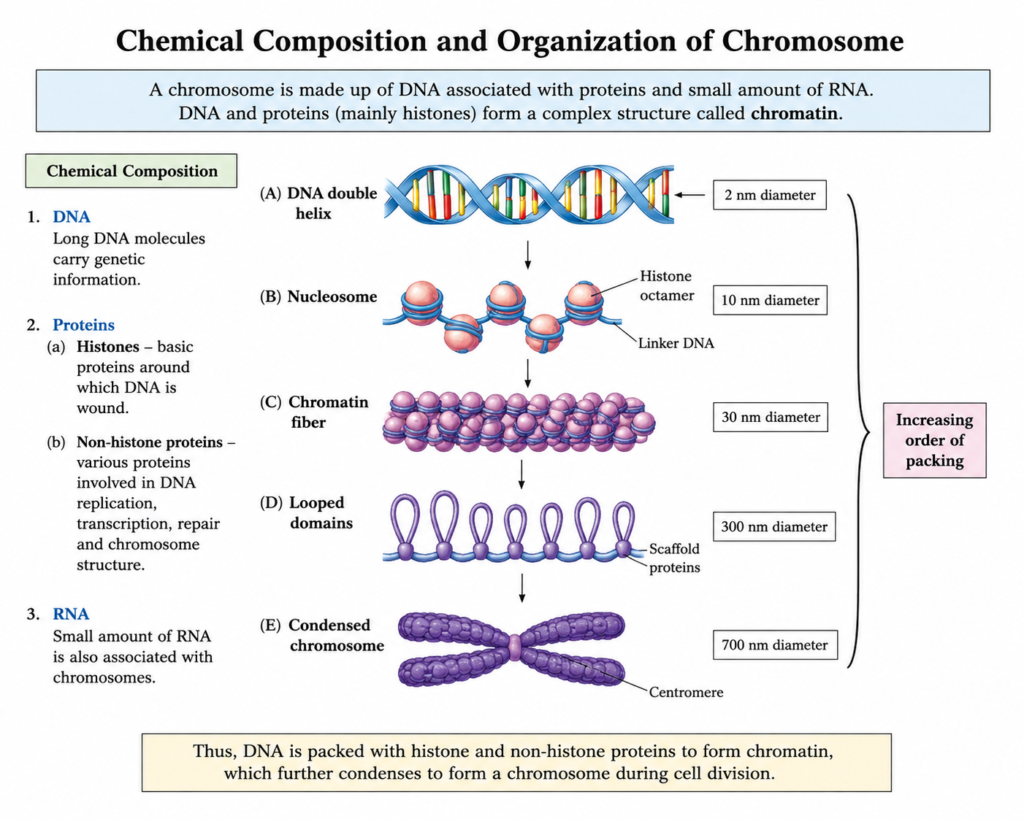
Chemical Composition of Chromosomes
Chromosomes are primarily composed of:
1. DNA
DNA (deoxyribonucleic acid) is the hereditary material of most organisms. It contains the nucleotide sequences that form genes as well as many regulatory and structural regions.
DNA provides the information required for:
- production of functional RNAs and proteins,
- regulation of cellular activities,
- DNA replication,
- inheritance of genetic information,
- and transmission of information during cell division.
2. Proteins
Chromosomal proteins can broadly be divided into histone and non-histone proteins.
Histones are basic proteins around which DNA is wrapped. They form the core of nucleosomes and play a major role in the packaging and regulation of chromosomal DNA. A nucleosome contains DNA wrapped around an octamer of core histones consisting of two copies each of H2A, H2B, H3, and H4.
Non-histone proteins include many proteins involved in DNA replication, transcription, repair, chromosome organization, condensation, and segregation.
3. RNA
RNA can also be associated with chromosomes. Chromosome-associated RNAs may participate in chromatin organization and gene regulation. However, DNA and proteins constitute the principal structural components of chromosomes.
Structure of a Chromosome
The appearance of a chromosome depends strongly on the stage of the cell cycle. A chromosome is relatively decondensed during much of interphase but becomes highly condensed during cell division, particularly around metaphase. This condensation makes individual chromosomes easier to observe and distinguish under a light microscope.
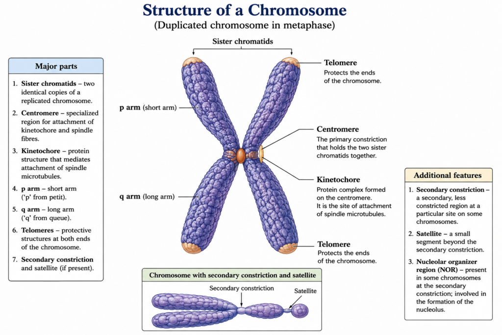
A typical duplicated metaphase chromosome has several recognizable features.
1. Sister Chromatids
After DNA replication, a chromosome consists of two sister chromatids. Each chromatid contains one complete DNA molecule associated with proteins.
The sister chromatids are genetically very similar because they arise from replication of the same chromosome. They remain associated until they are separated during chromosome segregation.
It is important to distinguish between a chromosome and a chromatid. Before DNA replication, a chromosome consists of one chromatid. After DNA replication, it consists of two sister chromatids that are still counted as one chromosome because they remain connected at the centromere.
2. Centromere
The centromere is a specialized chromosomal region that plays a central role in the accurate segregation of chromosomes during cell division.
In a condensed duplicated chromosome, the centromere appears as a constricted region where the sister chromatids remain associated. A specialized protein structure called the kinetochore forms at the centromeric region and interacts with spindle microtubules.
The major functions of the centromere include:
- maintaining the association of sister chromatids before their separation,
- providing the site for kinetochore assembly,
- helping chromosomes interact with the mitotic or meiotic spindle,
- and ensuring accurate chromosome segregation.
Errors in chromosome segregation can result in daughter cells receiving abnormal numbers of chromosomes.
3. Kinetochore
The kinetochore is a specialized protein complex assembled on the centromeric region of a chromosome.
During cell division, spindle microtubules attach to the kinetochore. This attachment allows chromosomes to interact with the spindle and move toward the appropriate daughter cells.
Thus, the centromere and kinetochore are closely related but are not identical terms:
- Centromere: specialized chromosomal region containing DNA and associated proteins.
- Kinetochore: protein complex assembled at the centromere that interacts with spindle microtubules.
This distinction is important when describing chromosome structure accurately.
4. Short Arm and Long Arm
The centromere divides a chromosome into two arms.
- The short arm is designated p.
- The long arm is designated q.
The letters come from the conventional cytogenetic terminology in which p refers to the short arm and q to the long arm.
The relative length of these arms depends on the position of the centromere.
5. Telomeres
Telomeres are specialized DNA-protein structures located at the ends of linear chromosomes.
They perform important protective and maintenance functions. Telomeric DNA helps distinguish natural chromosome ends from DNA breaks and contributes to the proper replication and stability of chromosome termini.
Without functional telomeres, chromosome ends can become unstable and may be incorrectly recognized as damaged DNA.
In humans, telomeres contain repeated DNA sequences with the repeat motif TTAGGG on the G-rich strand.
6. Secondary Constrictions
Some chromosomes contain additional constricted regions apart from the primary constriction represented by the centromere. These are called secondary constrictions.
A secondary constriction may correspond to a region associated with the formation of the nucleolus and ribosomal RNA genes. Such chromosome regions are commonly referred to as nucleolar organizer regions (NORs) when they contain clusters of ribosomal RNA genes that contribute to nucleolus formation.
The presence and position of secondary constrictions vary among species.
7. Satellite
A satellite is a small chromosome segment separated from the main body of a chromosome by a secondary constriction.
Chromosomes bearing such a segment are called satellite chromosomes.
The term “satellite” in this context refers to the visible structural segment of the chromosome and should not be confused with satellite DNA, which refers to repetitive DNA sequences.
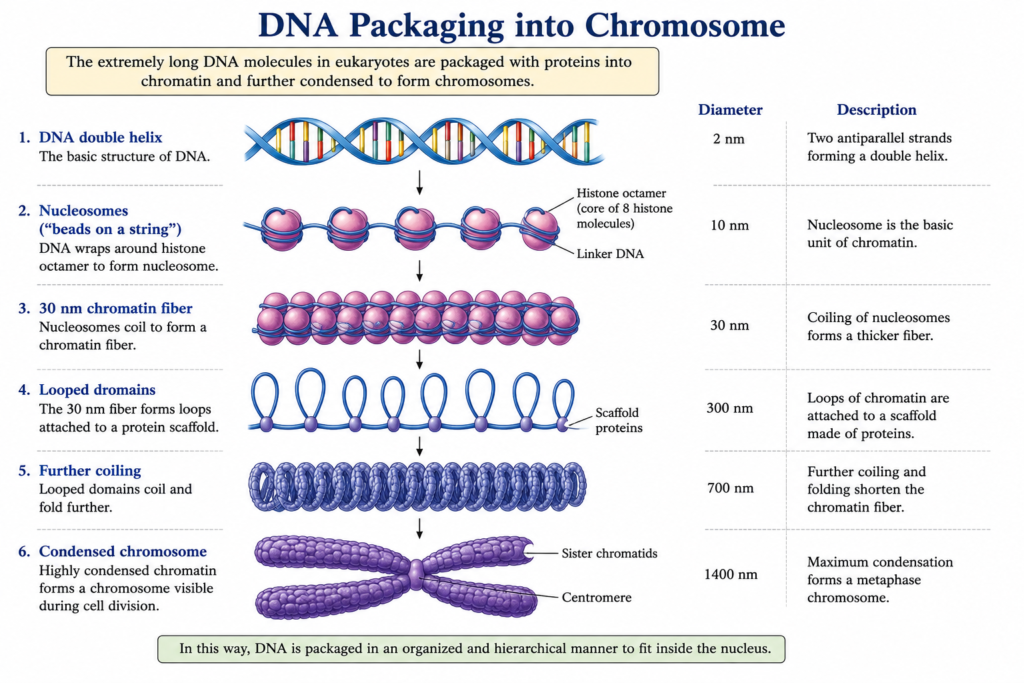
How Is DNA Packaged into a Chromosome?
One of the most remarkable features of chromosomes is their ability to package an enormous amount of DNA into a very small nuclear space.
The organization can be understood as a series of increasingly complex levels.
DNA double helix → nucleosomes → chromatin organization → loops and higher-order organization → condensed chromosome
At the first major level of packaging, DNA wraps around histone proteins to form nucleosomes. The nucleosomes are connected by linker DNA, producing the basic chromatin organization often described as a “beads-on-a-string” arrangement.
Further interactions and folding organize chromatin into larger structural domains. During mitosis, chromosome condensation becomes especially pronounced, producing the compact chromosomes that are commonly illustrated in textbooks.
Important clarification
Older textbooks sometimes describe chromosome packaging as a simple sequence of rigid structures, such as a fixed “30-nm fiber” followed by progressively thicker fibers. Modern research indicates that chromatin organization is more dynamic and complex than this simplified model suggests. Chromosomes contain loops and interacting domains whose organization can change according to cellular conditions and gene activity.
For undergraduate study, the nucleosome remains the fundamental and well-established unit of chromatin organization.
Euchromatin and Heterochromatin
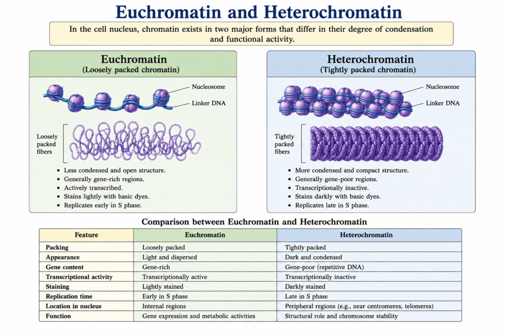
Chromatin is not uniformly packed throughout the nucleus. Two broad forms are commonly recognized:
Euchromatin
Euchromatin is relatively less condensed and is generally more accessible to the molecular machinery involved in gene transcription.
It tends to contain many actively expressed genes, although not every region of euchromatin is transcriptionally active at all times.
Heterochromatin
Heterochromatin is more highly condensed and generally less accessible to transcriptional machinery.
It is commonly associated with regions such as centromeres and chromosome ends, although heterochromatin can occur at many other chromosomal locations.
The distinction between euchromatin and heterochromatin is therefore related not only to how tightly DNA is packaged but also to its functional state and regulation.
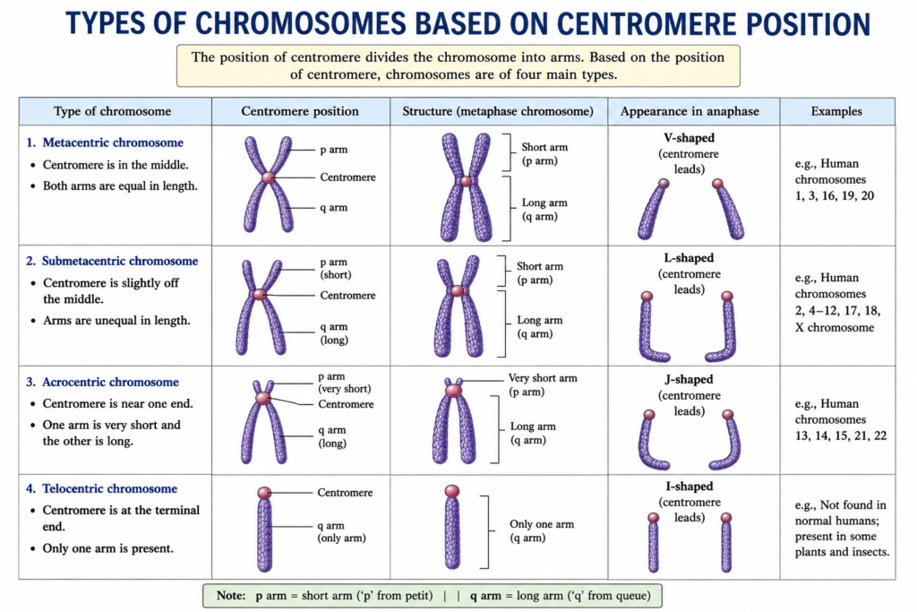
Types of Chromosomes Based on Centromere Position
Chromosomes can be classified according to the position of their centromere.
1. Metacentric Chromosome
In a metacentric chromosome, the centromere is approximately in the middle, producing two arms of nearly equal length.
During anaphase, such chromosomes commonly appear approximately V-shaped because of the movement of their centromeric regions toward the spindle poles.
2. Submetacentric Chromosome
In a submetacentric chromosome, the centromere is displaced from the middle, producing one shorter arm and one longer arm.
The chromosome may appear approximately L-shaped during anaphase.
3. Acrocentric Chromosome
In an acrocentric chromosome, the centromere lies close to one end, producing a very short p arm and a much longer q arm.
In humans, chromosomes 13, 14, 15, 21, and 22 are acrocentric chromosomes.
4. Telocentric Chromosome
In a telocentric chromosome, the centromere is located essentially at the terminal end, so only one major arm is apparent.
True telocentric chromosomes are found in some organisms but are not present in normal humans.
Functions of Chromosomes
Chromosomes perform several interconnected functions that are essential for cellular life and heredity.
1. Storage of Genetic Information
The primary function of chromosomes is to store genetic information in the form of DNA.
Genes are arranged along chromosomes in a linear order. These genes and their regulatory sequences provide information required for the production of functional molecules and the regulation of cellular processes.
2. Transmission of Hereditary Information
Chromosomes provide the physical basis for the transmission of genetic information from parent cells to daughter cells.
During mitosis, duplicated chromosomes are distributed between daughter cells. During meiosis, chromosome behavior contributes to the formation of genetically diverse gametes.
This chromosome behavior forms an important cellular foundation for heredity.
3. DNA Replication
Chromosomes must be replicated before cell division so that genetic information can be passed to daughter cells.
Eukaryotic chromosomes contain multiple origins of replication, allowing their very long DNA molecules to be copied efficiently.
4. Regulation of Gene Expression
Chromosome organization influences whether particular regions of DNA are accessible to transcription machinery.
Nucleosomes, histone modifications, DNA modifications, chromatin remodeling, and higher-order chromosome organization can all influence gene activity.
Thus, chromosomes are not simply passive containers for genes; their organization contributes to the regulation of genetic information.
5. Accurate Chromosome Segregation
The centromere and kinetochore are essential for the accurate movement and segregation of chromosomes during cell division.
Spindle microtubules interact with kinetochores, allowing chromosomes to be positioned and separated appropriately.
6. DNA Repair and Genome Stability
Chromosomes contain specialized regions and associated proteins that contribute to the maintenance of genome integrity.
Telomeres protect chromosome ends, while chromatin-associated proteins participate in DNA repair and chromosome organization.
7. Genetic Recombination
Chromosomes also participate in genetic recombination, particularly during meiosis.
The exchange of genetic material between homologous chromosomes can generate new combinations of alleles. This contributes to genetic variation within populations.
8. Organization of the Genome
Chromosomes divide the genome into distinct physical units.
Genes, regulatory sequences, repetitive DNA, structural regions, and other functional elements are organized along chromosomes. This organization helps the cell manage and regulate its genetic material.
Special Types of Chromosomes
Most chromosomes become highly condensed during cell division and can be studied as typical metaphase chromosomes. However, some chromosomes develop unusual structures that make them exceptionally large or visually distinctive.
Some organisms possess specialized chromosomes with unusual structures. Two important examples are lampbrush chromosomes and polytene chromosomes. Both are exceptionally large chromosomes and have been important in understanding chromosome organization and gene activity.
Lampbrush chromosomes are giant chromosomes characteristically observed in growing oocytes of many animals. They have an extended structure with prominent lateral loops associated with transcription.
Polytene chromosomes are giant chromosomes produced by repeated rounds of DNA replication without cell division. They are particularly well known in the salivary glands of dipteran insects and show characteristic banding patterns and transcription-associated puffs.
These are often called giant chromosomes because of their unusually large size compared with ordinary chromosomes.
Their unusual appearance is closely related to their developmental and cellular context.
Lampbrush Chromosomes
Lampbrush chromosomes are exceptionally large chromosomes found primarily in the growing oocytes of many animals, particularly amphibians and birds.
They become highly extended during a prolonged stage of meiotic prophase and display numerous lateral loops projecting from a central chromosomal axis. These loops are associated with active transcription, giving the chromosome its characteristic lampbrush-like appearance.
Lampbrush chromosomes are particularly valuable for studying the relationship between chromosome structure and transcription because actively transcribed regions can be visualized at a relatively large scale.
Main features
- Very large and highly extended chromosomes.
- Characteristically observed in growing oocytes.
- Possess a central axis with numerous lateral loops.
- Lateral loops are associated with active transcription.
- Useful for studying chromosome organization and gene activity.
Polytene Chromosomes
Polytene chromosomes are giant chromosomes produced when DNA replication occurs repeatedly without the corresponding cell divisions, resulting in many aligned copies of DNA within the same chromosome.
They are particularly well known from the salivary gland cells of dipteran insects, including Drosophila.
Polytene chromosomes show a characteristic pattern of alternating dark bands and lighter interbands. They are valuable for studying chromosome organization and gene activity because their large size and banding pattern make chromosomal regions comparatively easy to observe.
Certain regions may undergo localized decondensation, producing structures known as puffs, which are associated with increased transcriptional activity.
Main features
- Very large chromosomes produced through repeated DNA replication without cell division.
- Commonly studied in dipteran insects.
- Show characteristic bands and interbands.
- May contain transcriptionally active puff regions.
- Useful for studying chromosome structure, gene activity, and developmental regulation.
Importance of Chromosomes in Biology
Chromosomes provide the physical framework through which genetic information is organized and transmitted.
Their importance can be understood at several levels:
At the molecular level, chromosomes organize DNA with proteins and regulate access to genetic information.
At the cellular level, they ensure that replicated genetic material is distributed appropriately during cell division.
At the organismal level, chromosomes carry genes that contribute to development, physiology, reproduction, and inherited characteristics.
At the evolutionary level, chromosome structure, recombination, mutation, and changes in chromosome number or organization can influence genetic variation and evolution.
Chromosome analysis is also important in cytogenetics because changes in chromosome number or structure can be associated with genetic abnormalities. Chromosome size, number, morphology, and banding patterns can be examined using a karyotype or karyogram.
Chromosome Structure at a Glance
A typical duplicated chromosome can be remembered through the following arrangement:
Telomere → chromosome arm → centromere/kinetochore region → chromosome arm → telomere
After DNA replication:
One chromosome = two sister chromatids
Each chromatid contains:
DNA + histones + other chromosome-associated proteins
The DNA is organized as:
DNA → nucleosomes → chromatin → higher-order organization → condensed chromosome
This organization allows chromosomes to combine two apparently opposite requirements: compact packaging of enormous DNA molecules and controlled accessibility of that DNA when it is needed.
Key Takeaways
- A chromosome is a highly organized DNA–protein structure that carries genetic information.
- In eukaryotes, chromosomal DNA is associated with histones to form chromatin.
- A duplicated chromosome consists of two sister chromatids joined at the centromere.
- The centromere is essential for accurate chromosome segregation during cell division.
- The kinetochore is a protein complex assembled at the centromeric region and provides the attachment site for spindle microtubules.
- Telomeres protect the ends of linear chromosomes and contribute to chromosome stability.
- Chromosomes contain different regions of chromatin, including relatively open euchromatin and more condensed heterochromatin.
- Chromosomes help in DNA storage, replication, gene regulation, recombination, and transmission of hereditary information.
- Lampbrush chromosomes and polytene chromosomes are specialized giant chromosomes that provide unusual opportunities for studying chromosome organization and gene activity.
- The detailed study of lampbrush and polytene chromosomes is best treated separately because their structures and formation are considerably different from ordinary metaphase chromosomes.
Frequently Asked Questions
1. What is the main function of a chromosome?
The main function of a chromosome is to store and organize genetic information and ensure its accurate replication and transmission during cell division.
2. What is a chromosome made of?
A chromosome is primarily composed of DNA and proteins. Histones organize DNA into nucleosomes, while numerous non-histone proteins contribute to chromosome structure, replication, gene regulation, repair, and segregation.
3. What is the difference between a chromosome and chromatin?
Chromatin refers to the DNA-protein material in which eukaryotic DNA is organized. A chromosome is a discrete, highly organized unit of this chromatin containing a continuous DNA molecule.
4. What is the difference between a centromere and a kinetochore?
The centromere is a specialized chromosomal region, whereas the kinetochore is a protein complex assembled at the centromere that interacts with spindle microtubules.
5. What is the function of telomeres?
Telomeres help maintain and protect the ends of linear chromosomes and prevent chromosome ends from being incorrectly treated as DNA breaks.
6. Why are chromosomes condensed during cell division?
Condensation makes chromosomes more compact and mechanically manageable, helping their accurate movement and segregation during cell division.
7. What are lampbrush chromosomes?
Lampbrush chromosomes are giant chromosomes found mainly in growing oocytes. They contain prominent lateral loops associated with transcriptional activity.
8. What are polytene chromosomes?
Polytene chromosomes are giant chromosomes produced through repeated DNA replication without cell division. They are especially prominent in certain insect tissues and display characteristic banding patterns.
9. Are lampbrush and polytene chromosomes normal chromosomes?
Yes. They are specialized forms of chromosomes with unusual structural organization associated with particular developmental or cellular conditions.
10. Why are lampbrush and polytene chromosomes important?
Their large size and distinctive organization make them useful experimental systems for studying chromosome structure, DNA organization, transcription, and the relationship between chromatin structure and gene activity.
Conclusion
Chromosomes are much more than condensed structures visible during cell division. They are highly organized DNA-protein systems that coordinate the storage, replication, expression, protection, and transmission of genetic information.
Their structure includes specialized regions such as centromeres, kinetochores, chromosome arms, telomeres, and, in some chromosomes, secondary constrictions and satellite regions. At a molecular level, DNA is packaged with histones into nucleosomes and organized into increasingly complex chromatin structures. This organization is dynamic and allows the genome to remain compact while still providing controlled access to DNA.
Chromosomes also have essential roles in gene regulation, DNA replication, recombination, genome stability, and chromosome segregation.
Among the unusual forms, lampbrush and polytene chromosomes are especially important because their giant size and distinctive organization make chromosome activity easier to observe. Lampbrush chromosomes are characterized by prominent lateral loops, whereas polytene chromosomes arise from repeated DNA replication without cell division and display prominent bands and transcription-associated puffs.
Together, ordinary and specialized chromosomes demonstrate how efficiently biological systems organize enormous amounts of DNA while maintaining the ability to read, replicate, regulate, and transmit genetic information.
References
- Alberts B, Johnson A, Lewis J, et al. Molecular Biology of the Cell.
- Alberts B, Johnson A, Lewis J, et al. Molecular Biology of the Cell. Chromosomal DNA and Its Packaging in the Chromatin Fiber.
- Alberts B, Johnson A, Lewis J, et al. Molecular Biology of the Cell. The Global Structure of Chromosomes.
- Biology 2e. Chapter 10: Cell Division.
- Biology 2e. Chapter 13: Chromosomal Theory and Genetic Linkage.
- National Human Genome Research Institute. Chromosomes.
Discover more from Zoologyverse
Subscribe to get the latest posts sent to your email.