
1. Overview of Reproductive Hormones
Reproductive Hormones and Their Functions are central to the regulation of reproduction in animals. These chemical messengers coordinate the activities of the brain, pituitary gland and reproductive organs. They help control sexual development, gamete production, reproductive cycles and fertility.
The reproductive system depends on the coordinated action of several hormones. Some are released by the hypothalamus, while others are secreted by the pituitary gland, testes, ovaries and other reproductive tissues. Each hormone acts on specific target cells and produces a particular physiological response.
Reproductive Hormones and Their Functions are not limited to the production of sperm and eggs. They also influence sexual behaviour, pregnancy, birth and lactation in many animal groups. Their effects may vary with the animal’s sex, age, reproductive stage and environmental conditions.
The study of Reproductive Hormones and Their Functions helps explain how endocrine signals coordinate reproductive events. It also provides a foundation for understanding fertility, reproductive cycles and hormonal control in vertebrates.
In this article, Reproductive Hormones and Their Functions are examined through their sources, major types and roles in male and female reproduction.
2. Definition and General Characteristics of Reproductive Hormones
Reproductive hormones are chemical messengers produced by specialised endocrine cells and reproductive tissues. They travel through the bloodstream or act locally on nearby cells to regulate different reproductive processes. Reproductive Hormones and Their Functions involve the coordination of physiological activities necessary for sexual development and successful reproduction.
2.1 Definition of Reproductive Hormones
Reproductive hormones can be defined as biologically active chemical substances that regulate the development and functioning of the reproductive system. They control processes such as gametogenesis, sexual maturation, ovulation, reproductive cycles, pregnancy and lactation.
These hormones act by binding to specific receptors present on their target cells. This interaction activates cellular responses that produce particular reproductive effects.
2.2 General Characteristics of Reproductive Hormones
Reproductive hormones show several important characteristics:
- Chemical messengers: They transmit signals between endocrine glands and reproductive organs.
- Specific action: Each hormone acts on target cells containing suitable receptors.
- Blood transport: Many reproductive hormones travel through the bloodstream to reach distant target organs.
- Regulated secretion: Their release changes according to age, sex, reproductive stage and physiological requirements.
- Feedback regulation: Their secretion is controlled through positive and negative feedback mechanisms.
- Combined effects: Several hormones often work together to regulate a single reproductive process.
2.3 Importance of Hormonal Coordination
Reproductive Hormones and Their Functions depend on coordinated interactions between different endocrine organs. For example, pituitary gonadotropins regulate gonadal activity, while sex hormones influence reproductive development and provide feedback to the brain.
The actions of these hormones may differ among animal groups. Their secretion patterns also change during puberty, reproductive cycles, pregnancy and other reproductive stages.
Therefore, understanding Reproductive Hormones and Their Functions is essential for studying reproductive physiology and endocrine regulation in animals.
3. Classification of Reproductive Hormones
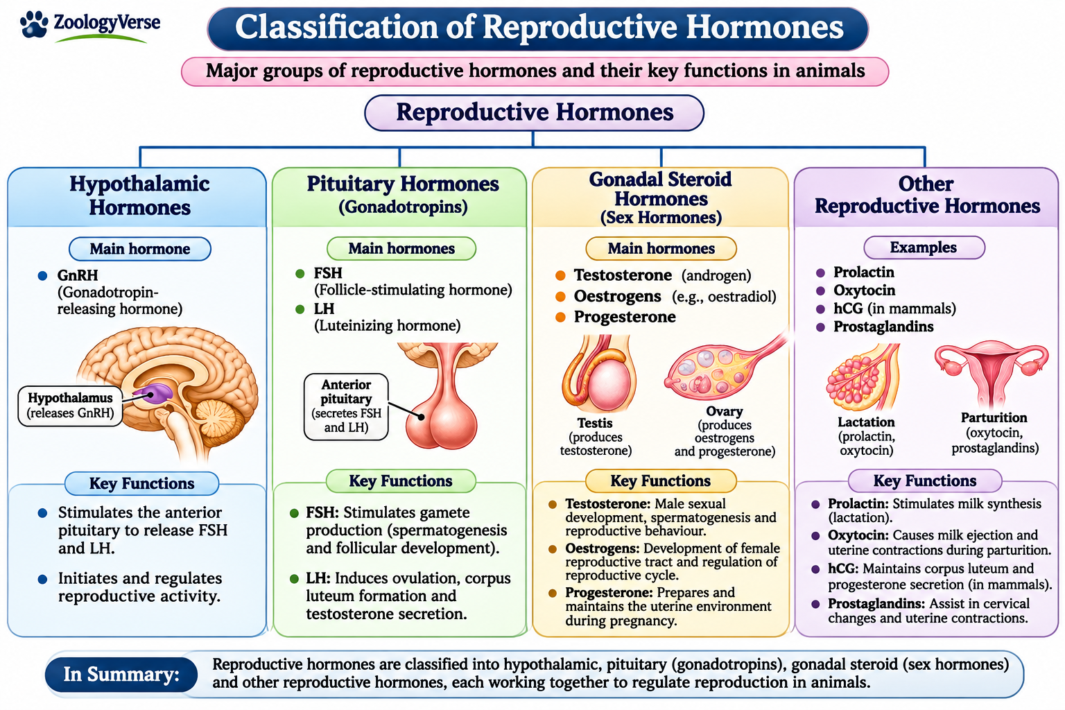
Reproductive Hormones and Their Functions can be classified according to their source, chemical nature and physiological roles. This classification helps explain how different hormones coordinate reproductive activities in animals.
Understanding Reproductive Hormones and Their Functions involves recognising four major groups: hypothalamic hormones, pituitary reproductive hormones, gonadal steroid hormones and other hormones involved in reproduction.
3.1 Hypothalamic Hormones
The hypothalamus produces gonadotropin-releasing hormone (GnRH). It stimulates the anterior pituitary gland to release FSH and LH. GnRH therefore initiates an important hormonal pathway controlling gonadal activity.
3.2 Pituitary Reproductive Hormones
The anterior pituitary secretes follicle-stimulating hormone (FSH) and luteinizing hormone (LH), collectively known as gonadotropins.
FSH supports sperm production and ovarian follicular development, while LH stimulates testosterone secretion in males and contributes to ovulation in females.
Prolactin also has reproductive roles, particularly in mammary gland development and milk production.
3.3 Gonadal Steroid Hormones
The testes and ovaries produce steroid hormones. The major examples are testosterone, oestrogens and progesterone.
These sex hormones regulate reproductive organ development, secondary sexual characteristics, reproductive cycles and pregnancy-related changes in animals.
3.4 Other Hormones Involved in Reproduction
Other important hormones include inhibin, activin, oxytocin, relaxin and anti-Müllerian hormone (AMH).
They contribute to the regulation of gonadotropin secretion, reproductive tract functions, childbirth, lactation and sexual differentiation.
This classification provides a framework for understanding Reproductive Hormones and Their Functions across different animal groups. Although these hormones belong to different groups, several interact to control the same reproductive event.
4. Hypothalamic Hormones – Gonadotropin-Releasing Hormone (GnRH)
The hypothalamus plays an important role in controlling reproductive hormone secretion. It produces gonadotropin-releasing hormone (GnRH), which regulates the activity of the anterior pituitary gland. Among Reproductive Hormones and Their Functions, GnRH is particularly important because it initiates the hormonal signals required for normal gonadal activity.
4.1 Source and Secretion of GnRH
GnRH is a peptide hormone produced by specialised neurosecretory neurons of the hypothalamus. These neurons are distributed in specific regions of the hypothalamus and send their nerve endings towards the median eminence.
GnRH is released into the hypophyseal portal blood circulation. This specialised vascular system transports the hormone directly to the anterior pituitary gland.
4.2 Structure of GnRH
GnRH is a small peptide hormone consisting of ten amino acids. It is therefore classified as a decapeptide hormone.
Its structure is highly conserved among vertebrates, although different animal groups may possess distinct molecular forms of GnRH. These variations are associated with differences in reproductive regulation among species.
4.3 Mechanism of Action
GnRH acts on specific receptors present on gonadotroph cells of the anterior pituitary gland. When it binds to these receptors, it activates intracellular signalling pathways.
This stimulation promotes the synthesis and secretion of two major gonadotropins:
- Follicle-Stimulating Hormone (FSH): Supports gamete development and follicular growth.
- Luteinizing Hormone (LH): Regulates gonadal steroid secretion and important reproductive events.
The released gonadotropins enter the bloodstream and act on the testes or ovaries. This hormonal pathway demonstrates the relationship between Reproductive Hormones and Their Functions.
4.4 Pulsatile Secretion of GnRH
GnRH is normally released in intermittent pulses rather than continuously. The frequency and amplitude of these pulses influence the secretion of FSH and LH.
Different pulse patterns can produce different pituitary responses. Continuous exposure to GnRH may initially stimulate gonadotropin release but can later reduce pituitary receptor responsiveness.
Therefore, pulsatile secretion is essential for maintaining normal reproductive hormone activity.
4.5 Role of GnRH in Male Reproduction
In males, GnRH stimulates the anterior pituitary gland to release FSH and LH.
FSH acts mainly on Sertoli cells and supports spermatogenesis. LH stimulates Leydig cells to produce testosterone.
Testosterone helps maintain male reproductive organs, secondary sexual characteristics and normal sperm production.
4.6 Role of GnRH in Female Reproduction
In females, GnRH regulates the secretion of FSH and LH, which control ovarian activity.
FSH stimulates ovarian follicular development, while LH contributes to final follicular maturation, ovulation and corpus luteum formation.
Changes in GnRH secretion patterns help coordinate hormonal changes during reproductive cycles. In many mammals, sustained high oestrogen levels before ovulation contribute to positive feedback and the preovulatory LH surge.
4.7 Regulation of GnRH Secretion
GnRH secretion is influenced by several physiological and environmental factors.
- Sex hormones: Testosterone, oestrogen and progesterone regulate GnRH secretion through feedback mechanisms.
- Nutritional status: Energy availability influences hypothalamic reproductive signalling.
- Stress: Increased stress hormone activity can suppress reproductive hormone secretion.
- Age: GnRH activity changes during development and sexual maturation.
- Environmental conditions: Photoperiod and seasonal signals influence reproductive activity in many animals.
These regulatory mechanisms help maintain hormonal coordination according to the reproductive requirements of each animal.
4.8 Biological Significance of GnRH
GnRH is essential for communication between the hypothalamus and anterior pituitary gland. It controls gonadotropin secretion and indirectly regulates gonadal development, gamete production and sex hormone synthesis.
Understanding GnRH is fundamental to studying Reproductive Hormones and Their Functions because it represents the initiating signal in the major hormonal pathway controlling vertebrate reproduction.
5. Pituitary Reproductive Hormones – FSH, LH and Prolactin
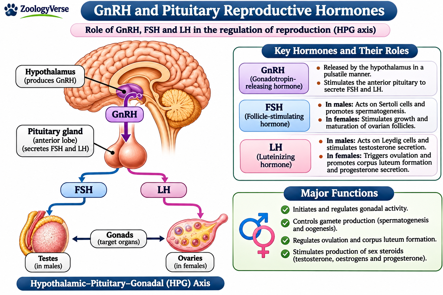
The anterior pituitary gland secretes several hormones that regulate reproductive activities in animals. Among them, follicle-stimulating hormone (FSH) and luteinizing hormone (LH) are the major gonadotropins. Prolactin also contributes to reproductive functions, particularly mammary gland development and lactation. These hormones represent an important group when studying Reproductive Hormones and Their Functions.
5.1 Follicle-Stimulating Hormone (FSH)
Follicle-stimulating hormone is a glycoprotein hormone secreted by gonadotroph cells of the anterior pituitary gland. Its secretion is stimulated by gonadotropin-releasing hormone (GnRH).
Functions of FSH:
- In males: FSH acts on Sertoli cells of the seminiferous tubules and supports sperm production.
- In females: It stimulates ovarian follicular growth and promotes oestrogen production by granulosa cells.
FSH therefore plays an essential role in gametogenesis and reproductive development.
5.2 Luteinizing Hormone (LH)
Luteinizing hormone is another glycoprotein gonadotropin released by the anterior pituitary gland.
Functions of LH:
- In males: It stimulates Leydig cells of the testes to produce testosterone.
- In females: It supports final follicular maturation, triggers ovulation in many mammals and promotes corpus luteum formation.
LH is particularly important for regulating gonadal steroid production and reproductive cycles.
5.3 Prolactin
Prolactin is a protein hormone secreted by lactotroph cells of the anterior pituitary gland. Unlike FSH and LH, it is not classified as a gonadotropin.
Functions of prolactin:
- Promotes mammary gland development.
- Stimulates milk synthesis after childbirth in mammals.
- Supports parental and reproductive behaviour in certain animal groups.
- Influences reproductive activity in some vertebrates.
The functions of these pituitary hormones demonstrate how Reproductive Hormones and Their Functions coordinate gamete production, sex hormone secretion and reproductive success.
5.4 Comparative Functions of Pituitary Reproductive Hormones
| Hormone | Main target | Major reproductive function |
|---|---|---|
| FSH | Sertoli cells and ovarian granulosa cells | Supports spermatogenesis and follicular development |
| LH | Leydig cells and ovarian follicular cells | Stimulates testosterone production and ovulation |
| Prolactin | Mammary glands and other target tissues | Promotes milk production and reproductive functions |
Together, these hormones connect hypothalamic signals with gonadal activity and other reproductive processes. Their coordinated secretion is an essential part of Reproductive Hormones and Their Functions in vertebrate animals.
6. Gonadal Steroid Hormones – Testosterone, Oestrogen and Progesterone
Gonadal steroid hormones are produced mainly by the testes and ovaries. They are lipid-soluble hormones derived from cholesterol. These hormones regulate sexual development, gametogenesis and pregnancy. Their actions form an important part of Reproductive Hormones and Their Functions because they affect reproductive organs and other tissues.
6.1 Testosterone
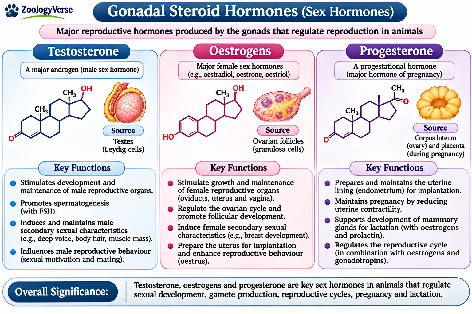
Testosterone is the principal androgen in most male vertebrates. It is produced mainly by Leydig cells in the testes under stimulation from luteinizing hormone (LH). Small amounts may also come from the ovaries and adrenal glands.
Testosterone supports the development and maintenance of male reproductive organs. It promotes secondary sexual characteristics, influences sexual behaviour and helps maintain spermatogenesis with follicle-stimulating hormone (FSH). These are major Reproductive Hormones and Their Functions in male animals.
6.2 Oestrogen
Oestrogens are steroid hormones produced mainly by ovarian follicles. Oestradiol is the major biologically active oestrogen in many female mammals. Smaller amounts are also produced by the testes, placenta and other tissues.
Oestrogen promotes female reproductive organ development and secondary sexual characteristics. It stimulates growth of the uterine lining and helps regulate ovarian cycles. In many mammals, sustained high oestrogen levels before ovulation produce positive feedback and contribute to the LH surge. These effects explain important Reproductive Hormones and Their Functions in female animals.
6.3 Progesterone
Progesterone is produced mainly by the corpus luteum after ovulation. During pregnancy, the placenta becomes an important source in many mammals.
Progesterone prepares the uterus for implantation by supporting the endometrium. It also helps maintain a suitable uterine environment during pregnancy and reduces uterine contractions in many species. Its changing levels regulate the luteal phase and contribute to the transition between reproductive cycles. These actions are central to Reproductive Hormones and Their Functions during pregnancy.
6.4 Comparative Functions of Gonadal Steroid Hormones
Testosterone, oestrogen and progesterone have distinct but interacting roles. Although testosterone is mainly associated with male reproductive development, all three hormones occur in both sexes. Their concentrations and effects vary with species, sex and reproductive stage. This interaction is an important feature of Reproductive Hormones and Their Functions.
| Hormone | Main source | Major reproductive functions |
|---|---|---|
| Testosterone | Leydig cells of testes | Male reproductive development, secondary sexual characteristics and support of spermatogenesis |
| Oestrogen | Ovarian follicles | Female reproductive development, uterine lining growth and regulation of reproductive cycles |
| Progesterone | Corpus luteum and placenta | Preparation and maintenance of the uterus for pregnancy |
The balanced secretion of these steroids coordinates sexual maturation, reproductive cycles and pregnancy. Understanding Reproductive Hormones and Their Functions therefore requires considering how these hormones act together, not only their individual effects.
7. Other Hormones Involved in Reproduction
Besides the major reproductive hormones, several additional hormones regulate important reproductive processes. They influence gonadotropin secretion, reproductive development, childbirth and lactation. Understanding these hormones provides a broader picture of Reproductive Hormones and Their Functions in animals.
7.1 Inhibin
Inhibin is a glycoprotein hormone produced mainly by Sertoli cells in males and granulosa cells in females.
Its principal function is to suppress follicle-stimulating hormone (FSH) secretion from the anterior pituitary gland. This selective negative feedback helps regulate spermatogenesis and ovarian follicular development.
Inhibin therefore contributes to hormonal balance within the reproductive system.
7.2 Activin
Activin is a protein hormone belonging to the transforming growth factor-beta (TGF-β) superfamily. It is produced by several tissues, including the gonads and pituitary gland.
Its major reproductive function is to stimulate FSH synthesis and secretion. Activin also participates in follicular development and reproductive tissue regulation.
Inhibin and activin have opposing effects on FSH secretion. Their interaction illustrates the complex nature of Reproductive Hormones and Their Functions.
7.3 Oxytocin
Oxytocin is a peptide hormone synthesised mainly by neurons of the hypothalamus and released into the bloodstream through the posterior pituitary gland.
Its major reproductive functions include:
- Stimulating uterine contractions during childbirth.
- Promoting milk ejection during lactation by causing contraction of myoepithelial cells surrounding mammary alveoli.
- Supporting reproductive and maternal behaviour in certain animal species.
Oxytocin is particularly important during parturition and lactation in mammals.
7.4 Relaxin
Relaxin is a peptide hormone produced mainly by the corpus luteum and placenta in many mammals. Its source and secretion patterns vary among animal species.
Its major functions include:
- Relaxing pelvic ligaments during pregnancy.
- Softening and remodelling the cervix before childbirth.
- Supporting changes in the reproductive tract during pregnancy.
- Contributing to maternal physiological adaptations.
Relaxin helps prepare the female reproductive system for parturition.
7.5 Anti-Müllerian Hormone (AMH)
Anti-Müllerian hormone is a glycoprotein hormone belonging to the TGF-β superfamily. It plays an important role in sexual differentiation and reproductive development.
In male embryos, AMH is secreted by Sertoli cells of the developing testes. It causes regression of the Müllerian ducts, which would otherwise develop into female internal reproductive structures.
In females, AMH is produced mainly by granulosa cells of developing ovarian follicles. It helps regulate follicular development and is also used as an indicator of ovarian reserve in several mammalian species.
7.6 Comparative Functions of Other Reproductive Hormones
| Hormone | Main source | Major function |
|---|---|---|
| Inhibin | Sertoli and granulosa cells | Suppresses FSH secretion |
| Activin | Gonads and other tissues | Stimulates FSH synthesis and secretion |
| Oxytocin | Hypothalamic neurons; released by posterior pituitary | Uterine contractions and milk ejection |
| Relaxin | Corpus luteum and placenta in many mammals | Supports pregnancy-related tissue changes and childbirth |
| AMH | Sertoli cells and ovarian granulosa cells | Müllerian duct regression and regulation of follicular development |
These hormones work alongside the major reproductive hormones to coordinate different stages of reproduction. Their combined actions demonstrate the diversity of Reproductive Hormones and Their Functions in animal physiology.
8. Functions of Reproductive Hormones in Male Animals
Male reproductive activity depends on coordinated hormonal signals from the hypothalamus, anterior pituitary gland and testes. Reproductive Hormones and Their Functions include the regulation of sexual maturation, sperm production, testosterone secretion and reproductive behaviour. These coordinated actions explain several important Reproductive Hormones and Their Functions in male animals.
8.1 Regulation of Sexual Maturation
At puberty, increased pulsatile secretion of gonadotropin-releasing hormone (GnRH) stimulates the anterior pituitary gland. It releases follicle-stimulating hormone (FSH) and luteinizing hormone (LH), which activate the testes.
Testosterone then supports the growth and maturation of male reproductive organs and the development of secondary sexual characteristics. This is one of the important Reproductive Hormones and Their Functions during male development. Reproductive Hormones and Their Functions also support the transition to reproductive maturity.
8.2 Regulation of Spermatogenesis
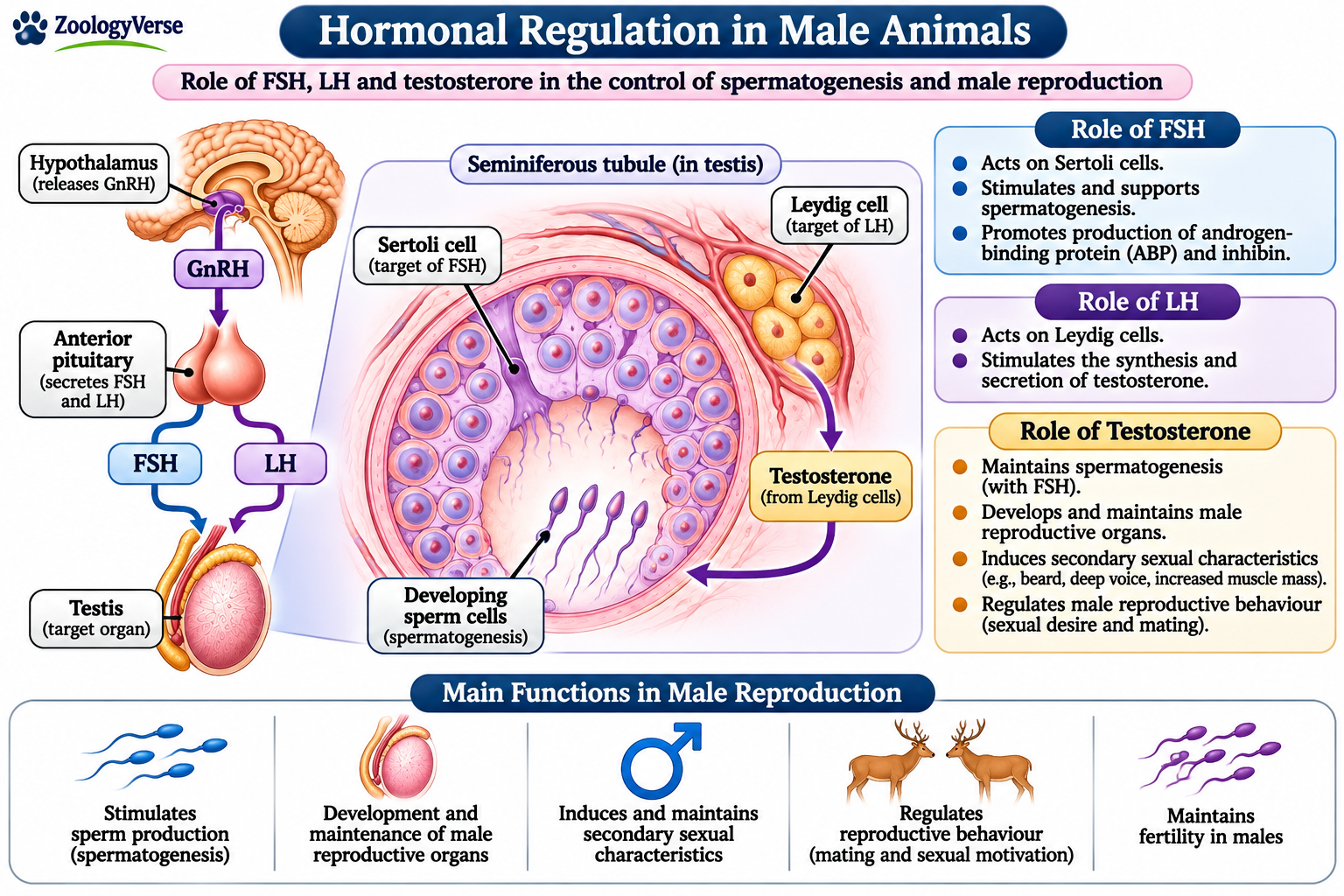
FSH acts mainly on Sertoli cells in the seminiferous tubules. These cells nourish and support developing germ cells. LH stimulates Leydig cells to produce testosterone, which acts within the testes and works with FSH to maintain spermatogenesis.
The combined action of these hormones supports the production and maturation of sperm. Therefore, Reproductive Hormones and Their Functions are essential for normal male fertility and reproductive capacity.
8.3 Regulation of Testosterone Secretion
LH controls testosterone production by stimulating Leydig cells. Testosterone supports reproductive organ function, libido and male secondary sexual characteristics.
It also provides negative feedback to the hypothalamus and pituitary gland, helping regulate further GnRH and LH secretion.
8.4 Regulation of Accessory Reproductive Glands and Behaviour
Testosterone supports the growth and activity of accessory reproductive glands, which contribute fluids to semen. It also influences sexual motivation and mating behaviour in many vertebrates, although behavioural responses vary among species.
8.5 Hormonal Feedback and Fertility
Testosterone provides feedback to the hypothalamus and pituitary, while inhibin from Sertoli cells selectively reduces FSH secretion. These mechanisms help maintain suitable hormone levels and testicular activity. They are another example of Reproductive Hormones and Their Functions working through feedback regulation.
Overall, Reproductive Hormones and Their Functions coordinate testicular development, sperm production, endocrine activity and reproductive behaviour. Their balanced secretion is necessary for normal male reproductive function and fertility.
9. Functions of Reproductive Hormones in Female Animals
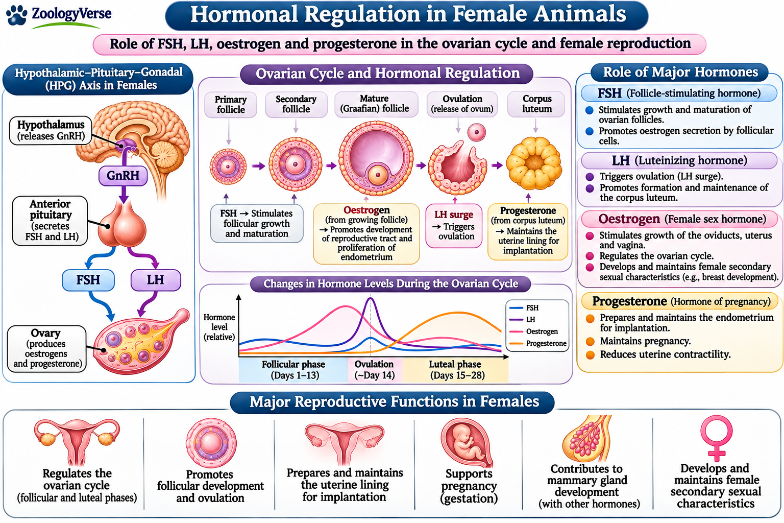
Female reproductive activity is regulated by interactions between the hypothalamus, anterior pituitary gland and ovaries. These organs produce hormones that coordinate ovarian development, ovulation, reproductive cycles and pregnancy. Reproductive Hormones and Their Functions are therefore essential for maintaining female fertility and reproductive health.
9.1 Regulation of Sexual Maturation
At puberty, increased secretion of gonadotropin-releasing hormone (GnRH) stimulates the anterior pituitary gland to release follicle-stimulating hormone (FSH) and luteinizing hormone (LH).
These gonadotropins activate the ovaries and promote follicular development. Oestrogen produced by developing follicles supports the maturation of female reproductive organs and the development of secondary sexual characteristics.
Thus, Reproductive Hormones and Their Functions play an important role in the transition from sexual immaturity to reproductive maturity.
9.2 Regulation of Ovarian Follicular Development
FSH stimulates the growth and development of ovarian follicles. It acts mainly on granulosa cells surrounding the developing oocyte.
These cells support follicular growth and produce oestrogen. LH also contributes to follicular development, particularly during the final stages of maturation.
The coordinated action of these hormones supports the development of mature follicles capable of releasing an oocyte.
9.3 Regulation of Ovulation
Ovulation is the release of a mature oocyte from an ovarian follicle. In many mammals, sustained high oestrogen levels before ovulation produce positive feedback on the hypothalamus and anterior pituitary gland.
This response leads to a sudden increase in LH secretion, known as the LH surge. It triggers follicular rupture and the release of the oocyte.
Reproductive Hormones and Their Functions are particularly important during this process because appropriate hormonal changes determine the timing of ovulation.
9.4 Regulation of the Corpus Luteum
After ovulation, the remaining follicular cells develop into the corpus luteum. This temporary endocrine structure secretes progesterone and, in many species, some oestrogen.
Progesterone prepares the uterine lining for possible implantation. It also helps maintain a suitable uterine environment during early pregnancy.
If pregnancy does not occur, the corpus luteum generally regresses in many mammals. The resulting decline in progesterone allows another reproductive cycle to begin.
9.5 Regulation of the Reproductive Cycle
Oestrogen and progesterone regulate changes in the female reproductive tract during reproductive cycles. Their concentrations rise and fall according to ovarian activity.
FSH supports follicular development, while LH contributes to ovulation and corpus luteum formation. These hormonal changes coordinate the different stages of the ovarian and oestrous cycles.
9.6 Support of Pregnancy and Parturition
Progesterone plays an important role in maintaining pregnancy by supporting the uterine environment and reducing uterine contractions in many mammals.
Oestrogens contribute to reproductive tract changes during pregnancy. Near childbirth, oxytocin stimulates uterine contractions, while relaxin supports changes in the cervix and pelvic tissues in several mammalian species.
These hormonal interactions demonstrate how Reproductive Hormones and Their Functions extend beyond ovulation to support successful reproduction.
9.7 Regulation of Lactation
Prolactin stimulates milk synthesis in mammary glands after childbirth. Oxytocin causes contraction of myoepithelial cells surrounding mammary alveoli, producing milk ejection.
Both hormones are important for successful lactation in mammals. Their actions support nourishment of newborn offspring.
9.8 Hormonal Feedback in Female Reproduction
Oestrogen and progesterone regulate hypothalamic and pituitary activity through feedback mechanisms. During much of the reproductive cycle, they generally produce negative feedback on GnRH, FSH and LH secretion.
However, sustained high oestrogen levels before ovulation produce positive feedback in many mammals. This leads to the preovulatory LH surge.
These feedback mechanisms maintain hormonal coordination and regulate reproductive events. They are a major aspect of Reproductive Hormones and Their Functions in female animals.
Overall, Reproductive Hormones and Their Functions coordinate sexual maturation, follicular development, ovulation, pregnancy and lactation. Their balanced secretion is essential for female reproductive success.
10. Hormonal Regulation During Reproductive Cycles
Reproductive cycles involve coordinated changes in the hypothalamus, pituitary gland and gonads. These changes regulate follicular development, ovulation, corpus luteum activity and reproductive readiness. Reproductive Hormones and Their Functions vary across the cycle, allowing reproductive events to occur in a coordinated sequence.
10.1 Role of the Hypothalamus and Pituitary Gland
The hypothalamus releases gonadotropin-releasing hormone (GnRH) in pulses. GnRH stimulates the anterior pituitary gland to secrete follicle-stimulating hormone (FSH) and luteinizing hormone (LH). This signalling sequence is a central part of Reproductive Hormones and Their Functions.
FSH promotes ovarian follicular growth, while LH supports follicular maturation, ovulation and corpus luteum formation. The frequency and amplitude of GnRH pulses influence gonadotropin secretion. These interactions form the foundation of Reproductive Hormones and Their Functions during reproductive cycles. The coordinated pituitary response illustrates how Reproductive Hormones and Their Functions control ovarian activity.
10.2 Hormonal Changes During the Follicular Phase
During the follicular phase of the ovarian cycle, FSH stimulates the growth of ovarian follicles. Developing follicles produce increasing amounts of oestrogen, which supports the growth and repair of the endometrium.
At moderate concentrations, oestrogen generally exerts negative feedback on GnRH and gonadotropin secretion. This helps regulate follicular development and prevents excessive stimulation. Reproductive Hormones and Their Functions therefore depend on changing hormone concentrations and feedback signals. This balance is central to Reproductive Hormones and Their Functions during follicular growth.
10.3 The Preovulatory LH Surge
As the dominant follicle matures, oestrogen secretion rises. When oestrogen remains at a sufficiently high level for an appropriate period, it produces positive feedback on the hypothalamus and pituitary gland in many mammals.
This positive feedback causes a sharp increase in LH secretion, called the LH surge. The surge initiates the final maturation of the oocyte and promotes follicular rupture, resulting in ovulation. This is a major example of Reproductive Hormones and Their Functions changing from negative to positive feedback. The LH surge demonstrates the precise timing required for Reproductive Hormones and Their Functions.
10.4 Hormonal Regulation During the Luteal Phase
After ovulation, the ruptured follicle develops into the corpus luteum under the influence of LH. The corpus luteum secretes progesterone and, in many species, oestrogen.
Progesterone prepares and maintains the uterine lining for possible pregnancy. This is one of the important reproductive outcomes governed by Reproductive Hormones and Their Functions. It also exerts negative feedback on GnRH, FSH and LH secretion, reducing the likelihood of another ovulation during the same cycle.
If fertilisation and pregnancy do not occur, the corpus luteum regresses in many mammals. Progesterone and oestrogen levels decline, allowing FSH secretion to rise and a new cycle to begin. Reproductive Hormones and Their Functions are thus closely linked to the formation and regression of the corpus luteum. This luteal activity is another essential component of Reproductive Hormones and Their Functions.
10.5 Hormonal Regulation of the Oestrous Cycle
In most non-primate mammals, reproductive activity follows an oestrous cycle. Its stages are commonly described as proestrus, oestrus, metestrus and diestrus, although the duration and hormonal patterns vary among species.
Oestrogen generally rises during follicular development and is associated with behavioural receptivity around oestrus. Progesterone becomes more prominent after ovulation when the corpus luteum is active. These changing levels coordinate reproductive behaviour and the condition of the reproductive tract. Their sequence highlights the role of Reproductive Hormones and Their Functions in the oestrous cycle.
10.6 Hormonal Regulation of the Menstrual Cycle
In primates, including humans, the menstrual cycle involves coordinated ovarian and uterine changes. The follicular phase is associated with follicular growth and rising oestrogen. After ovulation, the luteal phase is characterised by progesterone secretion from the corpus luteum.
If pregnancy does not occur, the corpus luteum degenerates. The decline in progesterone and oestrogen contributes to the shedding of the endometrium, known as menstruation. Reproductive Hormones and Their Functions regulate these cyclic changes through feedback between the ovaries, hypothalamus and pituitary gland. This ovarian-uterine coordination is a key feature of Reproductive Hormones and Their Functions.
10.7 Species Differences in Reproductive Cycles
Although the basic hormonal pathways are broadly conserved, reproductive cycles differ among animal groups. Some mammals are seasonal breeders, while others cycle throughout the year. The timing of ovulation may also differ; for example, some species are induced ovulators, in which mating stimulates the LH surge.
These differences show that Reproductive Hormones and Their Functions operate within species-specific reproductive patterns, influenced by physiology and environmental conditions. Such variation further illustrates the adaptability of Reproductive Hormones and Their Functions.
Overall, hormonal regulation coordinates the stages of reproductive cycles through changing secretion patterns and feedback mechanisms. The interaction of GnRH, FSH, LH, oestrogen and progesterone helps ensure that ovarian and uterine events remain appropriately synchronised.
11. Role of Reproductive Hormones in Pregnancy and Parturition
Pregnancy and parturition depend on coordinated hormonal interactions involving the ovaries, placenta, hypothalamus and pituitary gland. These hormones support the developing embryo or foetus, prepare maternal tissues and initiate childbirth. Reproductive Hormones and Their Functions change throughout gestation, with patterns that vary among animal species.
11.1 Hormonal Support During Early Pregnancy
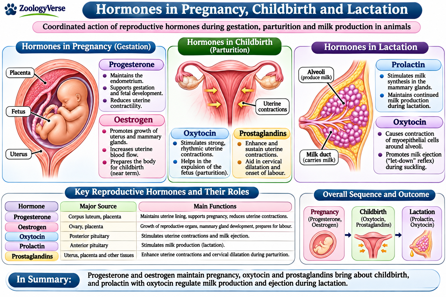
After ovulation, the corpus luteum secretes progesterone, which prepares the endometrium and supports a suitable uterine environment. In many mammals, progesterone reduces uterine contractility and helps maintain pregnancy. This is one of the central roles of Reproductive Hormones and Their Functions during early gestation, illustrating their importance in pregnancy maintenance.
In humans, human chorionic gonadotropin (hCG) maintains the corpus luteum during early pregnancy. Other mammals use different maternal-recognition signals to prevent corpus luteum regression. These species differences show that Reproductive Hormones and Their Functions are adapted to different reproductive systems. They also explain why Reproductive Hormones and Their Functions cannot be described by one identical pattern for all mammals.
11.2 Role of Progesterone and Oestrogens
Progesterone supports the endometrium, promotes uterine quiescence and helps regulate maternal tolerance of the developing conceptus. In some species, the corpus luteum remains the major progesterone source throughout gestation; in others, the placenta becomes a major source. Reproductive Hormones and Their Functions therefore involve different ovarian and placental contributions across mammals.
Oestrogens promote growth of the uterus and mammary glands and influence blood flow to reproductive tissues. Near the end of gestation, increased oestrogen activity can make the uterus more responsive to contraction signals. This changing hormonal balance is an important part of Reproductive Hormones and Their Functions during late pregnancy.
11.3 Endocrine Role of the Placenta
In many mammals, the placenta acts as an endocrine organ as well as an organ of exchange. Depending on the species, it produces progesterone, oestrogens, placental lactogen or chorionic gonadotropins.
Placental hormones help regulate maternal metabolism, support foetal growth and prepare mammary tissues for lactation. These actions broaden the contribution of Reproductive Hormones and Their Functions beyond the ovaries. The placenta therefore becomes an important contributor to Reproductive Hormones and Their Functions during pregnancy.
11.4 Hormonal Initiation of Parturition
Parturition is the process of giving birth. Its onset involves signals from the foetus, placenta and mother, and the precise mechanism differs among species. In sheep and several other domestic mammals, increased foetal cortisol helps trigger placental changes that promote prostaglandin production and labour.
Prostaglandins, particularly PGF₂α and PGE₂, contribute to cervical ripening and uterine contractions. In several domestic species, PGF₂α also causes corpus luteum regression, reducing progesterone support. These coordinated changes illustrate Reproductive Hormones and Their Functions in the transition from pregnancy to labour.
11.5 Role of Oxytocin and Relaxin
Oxytocin, released from the posterior pituitary gland, stimulates contractions of uterine smooth muscle. Cervical stretching during labour can promote further oxytocin release through a positive-feedback mechanism called the Ferguson reflex.
Relaxin contributes to cervical softening and relaxation of pelvic ligaments in several mammalian species. Its source and importance vary among animals. Alongside prostaglandins and oxytocin, it helps prepare the reproductive tract for delivery. These are important Reproductive Hormones and Their Functions at parturition. Together, they show how Reproductive Hormones and Their Functions coordinate the final stages of gestation.
11.6 Hormonal Coordination During Childbirth
Successful parturition depends on the interaction of progesterone withdrawal or reduced progesterone action, increased oestrogen influence, prostaglandin production and oxytocin-mediated contractions. The relative timing and importance of these changes differ among species.
Overall, Reproductive Hormones and Their Functions extend from maintaining pregnancy to preparing maternal tissues and coordinating labour. Their integrated actions support the safe delivery of offspring. This coordinated endocrine control is a defining feature of Reproductive Hormones and Their Functions during pregnancy and childbirth.
12. Role of Reproductive Hormones in Lactation
Lactation is the process of milk production and secretion by the mammary glands after childbirth. It provides nourishment and immune protection to newborn mammals. Reproductive Hormones and Their Functions are essential for initiating milk synthesis, maintaining milk production and controlling milk ejection.
12.1 Mammary Gland Development During Pregnancy
During pregnancy, oestrogens promote growth of the mammary ducts, while progesterone supports development of the lobules and alveoli. Prolactin, placental lactogen and other growth-related hormones contribute to mammary gland development and prepare the tissue for milk production.
Although these hormones support mammary development, their coordinated action is part of Reproductive Hormones and Their Functions. High concentrations of progesterone during pregnancy limit full milk secretion. After delivery, expulsion of the placenta causes a marked decline in progesterone and oestrogen. This change allows prolactin to stimulate active milk synthesis. This transition illustrates Reproductive Hormones and Their Functions in preparing the mammary glands for lactation.
12.2 Role of Prolactin in Milk Production
Prolactin is secreted by the anterior pituitary gland. It acts on mammary alveolar cells and stimulates the synthesis of milk components, including proteins, lactose and lipids.
After childbirth, suckling by the offspring stimulates sensory nerves in the nipple. Signals reach the hypothalamus and reduce the inhibitory influence of dopamine on prolactin secretion. As a result, prolactin release increases and supports continued milk production. This is one of the key Reproductive Hormones and Their Functions in the postpartum period.
Reproductive Hormones and Their Functions therefore depend on the repeated suckling stimulus to maintain milk synthesis in many mammals.
12.3 Role of Oxytocin in Milk Ejection
Oxytocin is released from the posterior pituitary gland in response to suckling. It causes contraction of myoepithelial cells surrounding the mammary alveoli and ducts. These contractions move milk towards the nipple, producing the milk-ejection or let-down reflex. This action is another important example of Reproductive Hormones and Their Functions.
Milk synthesis and milk ejection are separate processes: prolactin mainly promotes milk production, whereas oxytocin is responsible for milk release. Understanding these separate processes clarifies Reproductive Hormones and Their Functions. Their complementary actions demonstrate Reproductive Hormones and Their Functions during lactation. In some mammals, visual, auditory or other cues associated with offspring can also stimulate oxytocin release.
12.4 Suckling and Neuroendocrine Regulation
Suckling activates a neuroendocrine reflex involving sensory nerves, the hypothalamus and pituitary gland. It promotes prolactin secretion by reducing hypothalamic dopamine inhibition and stimulates oxytocin release from the posterior pituitary.
Frequent suckling generally helps maintain milk production and repeated milk ejection. In this way, Reproductive Hormones and Their Functions respond dynamically to the feeding activity of offspring. If suckling stops, prolactin stimulation decreases and milk production gradually declines. This feedback between offspring behaviour and maternal endocrine activity is a major example of Reproductive Hormones and Their Functions.
12.5 Other Hormones Supporting Lactation
Insulin, cortisol and thyroid hormones support the metabolic processes required for milk synthesis. Growth hormone can also contribute to mammary function, particularly in some domestic mammals. Their effects complement those of prolactin and oxytocin.
The relative importance of individual hormones differs among species and stages of lactation. These variations are also reflected in Reproductive Hormones and Their Functions. Therefore, Reproductive Hormones and Their Functions operate as an integrated endocrine system rather than through the action of a single hormone.
12.6 Biological Importance of Hormonal Control of Lactation
Hormonal regulation ensures that milk is produced and released in response to the needs of newborn offspring. Prolactin supports milk synthesis, while oxytocin enables milk ejection. These complementary roles summarise Reproductive Hormones and Their Functions in mammalian feeding. Other hormones maintain the metabolic and physiological conditions needed by the mammary glands. This coordinated support is essential to Reproductive Hormones and Their Functions throughout lactation.
Overall, Reproductive Hormones and Their Functions are central to successful lactation, linking maternal physiology with the nourishment and survival of offspring.
13. Comparative Overview of Reproductive Hormones in Animals
Reproduction in animals is regulated by hormones that coordinate sexual development, gamete production, reproductive cycles and parental functions. Although many endocrine signals are shared, their roles and patterns differ among animal groups.
Comparing Reproductive Hormones and Their Functions helps explain both conserved mechanisms and evolutionary adaptations. This comparison also shows how Reproductive Hormones and Their Functions are modified across animal lineages.
13.1 Common Hormonal Pathways in Vertebrates
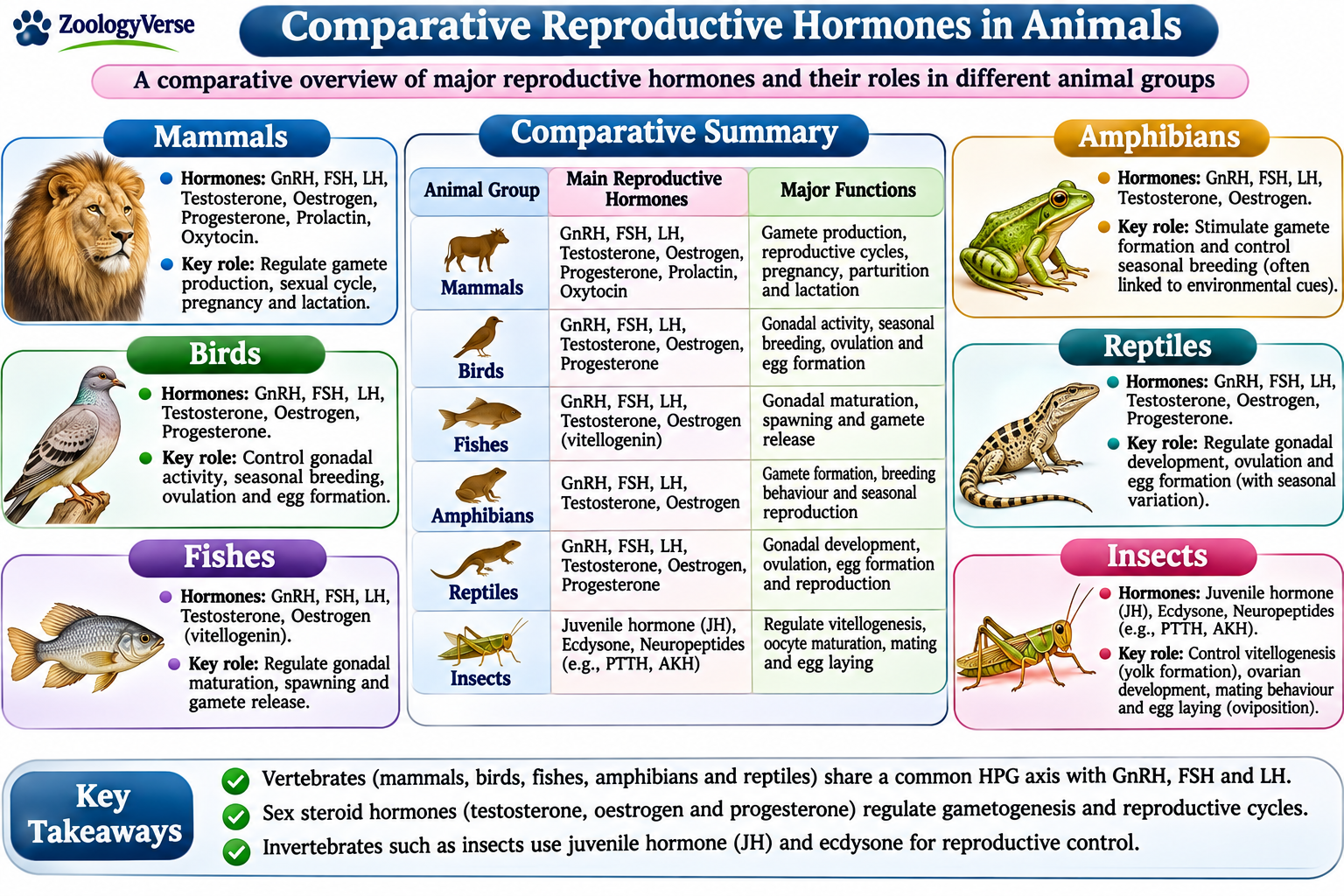
In most vertebrates, the hypothalamic-pituitary-gonadal (HPG) axis regulates reproductive activity. The hypothalamus releases gonadotropin-releasing hormone (GnRH), which stimulates the anterior pituitary gland to secrete follicle-stimulating hormone (FSH) and luteinizing hormone (LH).
These gonadotropins act on the ovaries or testes. They regulate gamete development and stimulate the production of sex steroids. This shared pathway is a central feature of Reproductive Hormones and Their Functions in vertebrates.
Testosterone, oestrogens and progesterone influence reproductive development and activity. Their effects depend on sex, species and reproductive stage.
The interaction between gonadotropins and gonadal steroids illustrates how Reproductive Hormones and Their Functions are coordinated through feedback mechanisms. These feedback systems are essential to Reproductive Hormones and Their Functions in regulating gonadal activity.
13.2 Reproductive Hormones in Mammals
In mammals, FSH and LH regulate ovarian follicular development, ovulation and testicular function. Testosterone supports male reproductive development and spermatogenesis. Oestrogens and progesterone regulate female reproductive cycles and help prepare the reproductive tract for pregnancy.
Prolactin stimulates milk production, while oxytocin causes milk ejection. During pregnancy, the placenta also produces hormones that support maternal and foetal physiology.
These processes demonstrate the wide range of Reproductive Hormones and Their Functions across mammalian life stages. The hormonal support of pregnancy and lactation further expands Reproductive Hormones and Their Functions beyond gamete production.
Hormonal patterns differ between mammalian species. In some animals, the corpus luteum is the main source of progesterone throughout pregnancy; in others, the placenta becomes a major source.
These differences show that Reproductive Hormones and Their Functions are adapted to species-specific reproductive systems.
13.3 Reproductive Hormones in Birds
Birds possess a hypothalamic-pituitary-gonadal axis that regulates gonadal development and reproductive activity. GnRH stimulates the release of FSH and LH, which act on the ovaries and testes.
In females, oestrogens support development of the reproductive tract and stimulate the liver to produce yolk precursor proteins.
In many bird species, day length influences hypothalamic activity and gonadotropin secretion. This seasonal response is an example of Reproductive Hormones and Their Functions interacting with environmental cues.
It highlights the role of Reproductive Hormones and Their Functions in synchronising breeding with favourable seasons.
13.4 Reproductive Hormones in Fishes
In fishes, the HPG axis regulates gametogenesis, gonadal maturation and spawning. GnRH stimulates pituitary gonadotropin secretion, and these hormones act on the gonads.
Oestrogens, androgens and progestogens regulate reproductive development. In many teleost fishes, maturation-inducing steroids help control final oocyte maturation and ovulation.
These pathways demonstrate the diversity of Reproductive Hormones and Their Functions in aquatic vertebrates. They also show how Reproductive Hormones and Their Functions are adjusted to different spawning strategies.
13.5 Reproductive Hormones in Amphibians and Reptiles
In amphibians, gonadotropins regulate ovarian and testicular activity. Sex steroids influence gamete development and reproductive behaviour. Seasonal conditions, including rainfall and temperature in some species, can affect reproductive timing.
Reptiles also use pituitary gonadotropins and gonadal steroids to regulate reproduction. In many species, temperature and seasonal changes influence gonadal activity.
These environmental influences show how Reproductive Hormones and Their Functions vary among ectothermic vertebrates. Such variation is an important feature of Reproductive Hormones and Their Functions in animals living under changing environmental conditions.
13.6 Hormonal Regulation in Invertebrates
Invertebrates do not all possess the vertebrate HPG axis. Their reproductive processes are regulated by diverse neuropeptides, biogenic amines and other signalling molecules.
In insects, juvenile hormone and ecdysteroids are important regulators. Juvenile hormone can influence reproductive maturation and vitellogenin production, while ecdysteroids participate in ovarian development and egg maturation.
Molluscs and crustaceans also use group-specific neuroendocrine signals. Their systems differ from vertebrate endocrine pathways, illustrating the evolutionary diversity of Reproductive Hormones and Their Functions across the animal kingdom.
The comparison emphasises that Reproductive Hormones and Their Functions are not identical in all animals.
13.7 Comparative Summary
| Animal group | Important hormonal signals | Major reproductive roles |
|---|---|---|
| Mammals | GnRH, FSH, LH, testosterone, oestrogens, progesterone, prolactin and oxytocin | Gametogenesis, reproductive cycles, pregnancy and lactation |
| Birds | GnRH, FSH, LH and oestrogens | Gonadal activity, yolk formation and seasonal breeding |
| Fishes | GnRH, gonadotropins, oestrogens, androgens and progestogens | Gametogenesis, final maturation and spawning |
| Amphibians | Gonadotropins and sex steroids | Gonadal development, gamete production and seasonal reproduction |
| Reptiles | Gonadotropins and sex steroids | Gonadal activity and seasonal reproductive timing |
| Insects | Juvenile hormone, ecdysteroids and neuropeptides | Reproductive maturation, vitellogenesis and egg development |
| Other invertebrates | Group-specific neuropeptides and signalling molecules | Regulation of gamete development and reproductive behaviour |
13.8 Evolutionary Significance of Hormonal Diversity
Several major reproductive pathways are conserved among vertebrates. However, the timing and effects of hormonal signals have been modified according to different life histories, habitats and breeding patterns.
These similarities and differences help explain the evolution of Reproductive Hormones and Their Functions. Invertebrates show a wider range of endocrine arrangements. Their reproductive signals reflect distinct evolutionary histories and adaptations.
A comparative approach therefore helps distinguish broadly shared endocrine principles from mechanisms specific to particular animal groups. It clarifies how Reproductive Hormones and Their Functions support different reproductive modes.
Overall, Reproductive Hormones and Their Functions provide a framework for understanding how animals coordinate reproduction while adapting to different ecological conditions. Their study connects endocrinology with the diversity of reproductive strategies across the animal kingdom.
14. Factors Affecting Hormonal Secretion
Hormonal secretion is influenced by internal physiological processes and external environmental conditions. These factors affect the timing and amount of hormone release, thereby influencing reproductive activity.
Reproductive Hormones and Their Functions depend on coordinated regulation that maintains normal reproductive processes.
14.1 Age and Developmental Stage
Hormone secretion changes throughout an animal’s life. During puberty, increased hypothalamic and pituitary activity stimulates gonadal development.
In adulthood, reproductive hormone patterns support gamete production and reproductive cycles. Age-related changes may reduce gonadal activity and alter hormone concentrations.
These changes influence Reproductive Hormones and Their Functions at different stages of life. Developmental transitions therefore shape Reproductive Hormones and Their Functions over an animal’s lifetime.
14.2 Nutrition and Body Condition
Adequate nutrition is essential for normal reproductive activity. Energy deficiency, poor-quality diets and low body condition can suppress hypothalamic signalling and reduce gonadotropin secretion.
In females, inadequate nutrition may delay puberty, disturb ovarian cycles or reduce fertility. In males, prolonged nutritional deficiency can affect testosterone production and sperm development.
Thus, nutritional status is an important influence on Reproductive Hormones and Their Functions. Energy balance is closely linked to Reproductive Hormones and Their Functions in both sexes.
14.3 Environmental Factors and Photoperiod
Environmental conditions such as temperature, rainfall and day length can influence reproductive hormone secretion. In seasonal breeders, changes in photoperiod affect hypothalamic pathways and alter gonadotropin release.
These responses help synchronise reproduction with favourable environmental conditions. They demonstrate how Reproductive Hormones and Their Functions respond to environmental information.
14.4 Stress and Physiological Condition
Physical and psychological stress can activate the hypothalamic-pituitary-adrenal axis. Increased glucocorticoid secretion may interfere with the hypothalamic-pituitary-gonadal axis and suppress reproductive activity.
Severe illness, injury and prolonged stress may disturb reproductive cycles, ovulation and sperm production. Stress-related changes can alter Reproductive Hormones and Their Functions by affecting hormone release and feedback.
14.5 Feedback Mechanisms
Hormone secretion is regulated through negative and positive feedback. Gonadal hormones such as testosterone, oestrogen and progesterone act on the hypothalamus and pituitary gland to adjust GnRH and gonadotropin secretion.
Positive feedback occurs in specific situations, such as the preovulatory oestrogen surge in many mammals. These feedback systems maintain the balance required for Reproductive Hormones and Their Functions.
14.6 Seasonal and Biological Rhythms
Some animals show daily, monthly or seasonal rhythms in reproductive activity. These rhythms are influenced by biological clocks and environmental signals.
Seasonal changes can affect gonadal size, hormone concentrations and breeding behaviour. Such rhythms illustrate the flexibility of Reproductive Hormones and Their Functions across species.
14.7 Disease, Drugs and Endocrine Disruptors
Diseases affecting the hypothalamus, pituitary gland, thyroid gland or gonads may alter reproductive hormone secretion. Certain medicines can also influence hormone synthesis, release or action.
Endocrine-disrupting chemicals may interfere with hormone receptors or normal signalling pathways. Exposure can affect fertility and reproductive development in animals.
These influences are important when evaluating Reproductive Hormones and Their Functions in health and disease.
14.8 Species and Individual Differences
Hormonal patterns vary according to species, sex, age, reproductive status and genetic background. Even individuals of the same species may show differences related to health, nutrition and environmental exposure.
These variations mean that normal hormone concentrations and reproductive patterns must be interpreted in the context of the animal being studied. This context is essential when comparing Reproductive Hormones and Their Functions between species.
Overall, reproductive hormone secretion is shaped by interacting physiological, environmental and genetic factors. Their combined effects determine reproductive readiness, fertility and reproductive success.
15. Biological Significance of Reproductive Hormones
Reproduction is essential for the continuation of animal species. Hormones coordinate the physiological processes required for sexual development, gamete production, fertilisation, pregnancy and parental care. Reproductive Hormones and Their Functions therefore have a central role in reproductive success and species survival.
15.1 Regulation of Sexual Maturation
Reproductive hormones initiate and regulate puberty. GnRH stimulates the pituitary gland to release FSH and LH, which activate the gonads. Gonadal steroids then support the development of reproductive organs and secondary sexual characteristics.
These changes prepare animals for reproductive activity. They demonstrate how Reproductive Hormones and Their Functions connect endocrine development with sexual maturity. This developmental role is one foundation of Reproductive Hormones and Their Functions.
15.2 Support of Gamete Production
FSH and LH regulate important stages of gametogenesis in both sexes. In males, FSH acts on Sertoli cells, while LH stimulates Leydig cells to produce testosterone. These activities support sperm development and maturation.
In females, FSH promotes ovarian follicular development, while LH contributes to final follicular maturation and ovulation. This hormonal coordination is a major aspect of Reproductive Hormones and Their Functions because successful reproduction depends on the production of functional gametes. It highlights the importance of Reproductive Hormones and Their Functions in maintaining fertility.
15.3 Coordination of Reproductive Cycles
Hormonal changes regulate ovarian and uterine events during reproductive cycles. Oestrogen and progesterone influence the reproductive tract, while gonadotropins control ovarian activity.
In many mammals, these hormonal patterns also influence sexual receptivity and mating behaviour. Reproductive Hormones and Their Functions help synchronise reproductive physiology with the appropriate stages of the cycle.
15.4 Regulation of Fertilisation and Pregnancy
Hormones support conditions that make fertilisation and pregnancy possible. Oestrogens influence the female reproductive tract, while progesterone prepares and maintains the uterine environment in many mammals.
During pregnancy, placental and ovarian hormones support maternal physiology and foetal development. These roles show that Reproductive Hormones and Their Functions extend beyond gamete production to the maintenance of gestation. They further emphasise the importance of Reproductive Hormones and Their Functions throughout the reproductive lifespan.
15.5 Control of Parturition and Lactation
Near childbirth, hormonal changes help prepare the reproductive tract for delivery. Prostaglandins contribute to cervical changes and uterine contractions, while oxytocin stimulates uterine smooth muscle contractions.
After birth, prolactin stimulates milk synthesis and oxytocin promotes milk ejection. These processes illustrate Reproductive Hormones and Their Functions in supporting newborn nourishment and maternal care.
15.6 Influence on Reproductive Behaviour
Sex steroids can influence courtship, mating behaviour and sexual motivation in many animals. Their effects vary with species, sex, season and social conditions.
Hormonal signals can therefore connect reproductive physiology with behaviour. This relationship is another important part of Reproductive Hormones and Their Functions.
15.7 Adaptation to Environmental Conditions
Reproductive activity in many animals is influenced by environmental cues such as photoperiod, temperature, rainfall and food availability. Endocrine pathways help translate these cues into changes in gonadal activity and breeding timing.
Seasonal breeders, for example, may show changes in gonadotropin secretion and sex steroid concentrations across the year. This flexibility demonstrates how Reproductive Hormones and Their Functions contribute to reproductive adaptation.
15.8 Maintenance of Reproductive Health
Balanced hormone secretion is necessary for normal reproductive function. Disturbances in the hypothalamus, pituitary gland or gonads can affect fertility, gamete production and reproductive cycles.
Understanding Reproductive Hormones and Their Functions is therefore important in animal breeding, veterinary medicine, reproductive research and the management of threatened species. Hormonal knowledge can help identify reproductive disorders and support appropriate breeding programmes.
Overall, Reproductive Hormones and Their Functions coordinate multiple processes that contribute to successful reproduction. Their actions link sexual development, fertility, pregnancy, birth and parental care, making them essential to the continuation of animal populations.
16. Conclusion
Reproductive Hormones and Their Functions are essential to the coordination of animal reproduction. Hormones released by the hypothalamus, pituitary gland and gonads regulate sexual maturation, gamete production and reproductive cycles. Their actions ensure that reproductive events occur in an organised and coordinated manner.
The hypothalamic-pituitary-gonadal axis provides the central regulatory pathway in many vertebrates. GnRH stimulates the secretion of FSH and LH, which act on the gonads to regulate gametogenesis and steroid hormone production. Testosterone, oestrogens and progesterone then influence reproductive development, cycles and reproductive behaviour. These interactions explain the importance of Reproductive Hormones and Their Functions in both male and female animals.
During pregnancy, progesterone and other maternal and placental hormones support the developing embryo or foetus. Near parturition, prostaglandins, oxytocin and other signals contribute to the processes involved in childbirth. After delivery, prolactin and oxytocin regulate milk production and ejection. These coordinated activities demonstrate that Reproductive Hormones and Their Functions extend beyond fertilisation to include pregnancy, birth and parental care.
Hormonal secretion is affected by age, nutrition, environmental conditions, stress, disease and biological rhythms. Such influences can alter reproductive timing and fertility. In addition, hormonal pathways differ among animal groups, reflecting their evolutionary histories and reproductive adaptations. Understanding Reproductive Hormones and Their Functions is therefore important in comparative zoology, animal breeding, veterinary medicine and wildlife conservation.
In conclusion, Reproductive Hormones and Their Functions provide the endocrine coordination required for successful reproduction. Their balanced activity supports reproductive health, fertility and the continuation of animal populations.
17. Frequently Asked Questions (FAQs)
What are reproductive hormones?
Which hormones control reproduction in animals?
What is the role of GnRH in reproduction?
What are the functions of FSH and LH?
What is the role of progesterone during pregnancy?
Which hormones are responsible for milk production and ejection?
How do environmental factors affect reproductive hormones?
Do all animals have the same reproductive hormones?
What happens when reproductive hormone levels become abnormal?
18. References
- Marques, P., De Sousa Lages, A., Skorupskaite, K., Rozario, K. S., Anderson, R. A., and George, J. T. (2024). Physiology of GnRH and Gonadotrophin Secretion. Endotext. NCBI Bookshelf.
https://www.ncbi.nlm.nih.gov/books/NBK279070/ - Plant, T. M., and Zeleznik, A. J. (Eds.). (2015). Knobil and Neill’s Physiology of Reproduction (4th ed.). Academic Press.
- Endocrinology of the Testis and Spermatogenesis. Endotext. NCBI Bookshelf.
https://www.ncbi.nlm.nih.gov/books/NBK279031/ - Hormonal Regulation of Female Reproduction. (2013). Cold Spring Harbor Perspectives in Biology.
https://pmc.ncbi.nlm.nih.gov/articles/PMC3647363/ - Schuler, G., Fürbass, R., and Klisch, K. (2018). Placental contribution to the endocrinology of gestation and parturition. Animal Reproduction, 15(Supplement 1), 822–842.
https://doi.org/10.21451/1984-3143-AR2018-0015 - Hamburg-Shields, E., and Mesiano, S. (2024). The hormonal control of parturition. Physiological Reviews, 104(3).
https://doi.org/10.1152/physrev.00019.2023 - Hannan, F. M., Elajnaf, T., Vandenberg, L. N., Kennedy, S. H., and Thakker, R. V. (2023). Hormonal regulation of mammary gland development and lactation. Nature Reviews Endocrinology, 19, 46–61.
https://doi.org/10.1038/s41574-022-00742-y - Origin and Evolution of the Neuroendocrine Control of Reproduction in Vertebrates, With Special Focus on Genome and Gene Duplications. (2020). Physiological Reviews.
https://doi.org/10.1152/physrev.00009.2019 - Riddiford, L. M. (2012). How does juvenile hormone control insect metamorphosis and reproduction? General and Comparative Endocrinology, 179(3), 477–484.
https://doi.org/10.1016/j.ygcen.2012.06.001 - Van Wielendaele, P., Badisco, L., and Vanden Broeck, J. (2013). Neuropeptidergic regulation of reproduction in insects. General and Comparative Endocrinology, 188, 23–34.
https://doi.org/10.1016/j.ygcen.2013.02.005 - Khalid, M. Z., Ahmad, S., Ngegba, P. M., and Zhong, G. (2021). Role of Endocrine System in the Regulation of Female Insect Reproduction. Biology, 10(7), 614.
https://doi.org/10.3390/biology10070614
Discover more from Zoologyverse
Subscribe to get the latest posts sent to your email.