
Gametogenesis: Spermatogenesis and Oogenesis Explained
Every human being starts life as a single cell — and that cell only exists because two highly specialized cells, a sperm and an egg, managed to find each other and fuse. But sperm and eggs aren’t just “made” the way skin cells or blood cells are made. They’re produced through a carefully choreographed process called gametogenesis, and the male and female versions of this process are surprisingly different from each other, even though they’re solving the same basic problem: turning a regular diploid cell into a haploid gamete.
In this article, we’ll walk through what gametogenesis actually is, break down spermatogenesis and oogenesis stage by stage, and then line them up side by side so the differences are easy to remember — whether you’re studying for an exam or just curious about how human reproduction works at the cellular level.
What Is Gametogenesis?
Gametogenesis is the biological process by which specialized germ cells develop into mature gametes — sperm in males and eggs (ova) in females. The whole point of this process is chromosome reduction: a normal body cell in humans is diploid, meaning it carries 46 chromosomes (23 pairs), but a gamete needs to be haploid, carrying only 23 chromosomes. That way, when a sperm and egg fuse during fertilization, the resulting embryo ends up with the correct 46 chromosomes total — half from each parent.
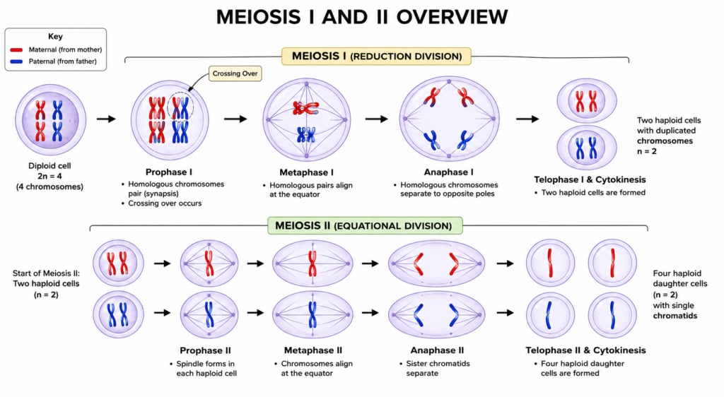
This chromosome halving happens through meiosis, a special type of cell division involving two consecutive rounds of division (meiosis I and meiosis II) that follow a single round of DNA replication. Meiosis is the shared engine behind both spermatogenesis and oogenesis, but how each process uses that engine — and what it produces at the end — is where things start to diverge.
Gametogenesis is split into two parallel pathways:
- Spermatogenesis — the formation of sperm cells in the testes
- Oogenesis — the formation of egg cells in the ovaries
A Quick Refresher on Meiosis
Since both pathways depend on it, it’s worth being precise about what meiosis actually does. A germ cell first duplicates its DNA so that each of its 46 chromosomes now consists of two identical sister chromatids. In meiosis I, homologous chromosome pairs (one from each parent) line up together, swap segments of DNA through a process called crossing over, and then separate — this is what actually cuts the chromosome number in half, from 46 to 23. In meiosis II, which follows without another round of DNA replication, the sister chromatids of each chromosome are pulled apart, similar to what happens in ordinary mitosis. The net result of one germ cell going through both divisions is four haploid daughter cells — though, as you’ll see, oogenesis manipulates this outcome so that only one of those four actually becomes usable.
This is also where genetic diversity comes from. Crossing over during meiosis I, combined with the random way maternal and paternal chromosomes get distributed into daughter cells (independent assortment), means no two sperm or eggs carry an identical set of chromosomes — which is why siblings look different from each other despite sharing the same parents.
Let’s look at each pathway individually before comparing them.
Spermatogenesis: How Sperm Cells Are Made
Spermatogenesis takes place in the seminiferous tubules of the testes, and it begins at puberty. Unlike oogenesis, it doesn’t start before birth — it kicks off once the body starts producing enough testosterone and other hormonal signals to activate the process, and then it continues more or less nonstop for the rest of a man’s life.
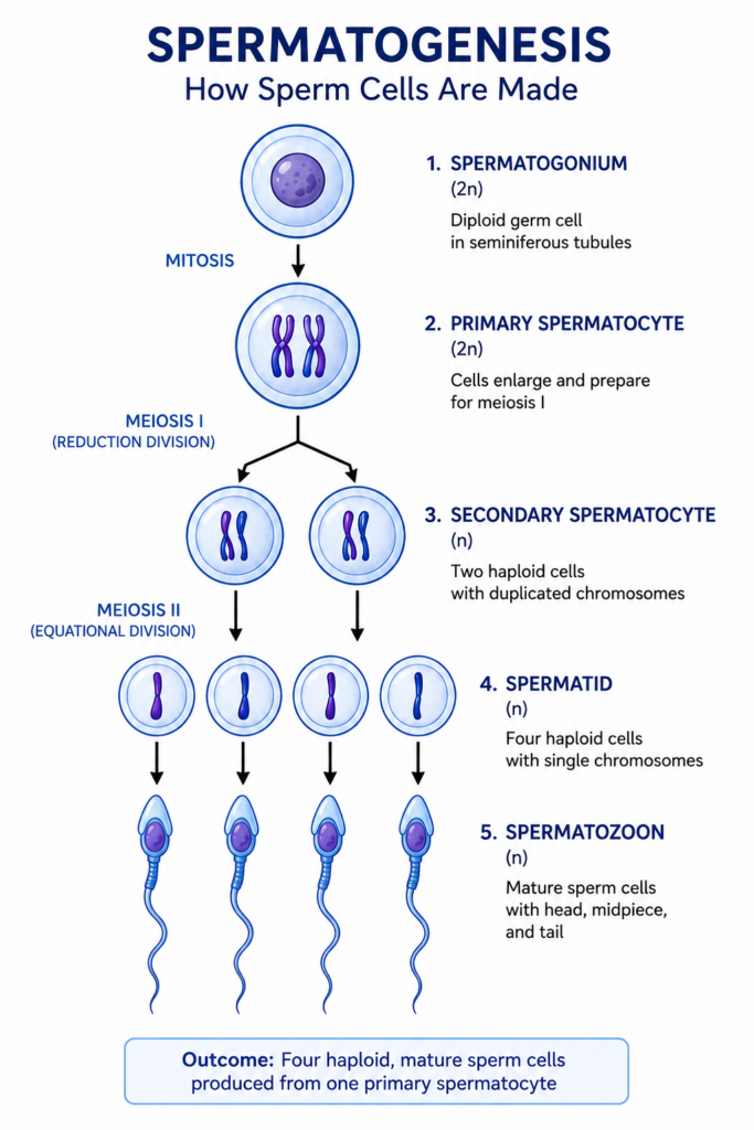
The stages of spermatogenesis are:
1. Spermatogonia (the starting germ cells). These diploid cells sit at the outer edge of the seminiferous tubules and act as a stem-cell reserve. Some divide by mitosis simply to keep replenishing this pool, while others commit to becoming sperm.
2. Primary spermatocytes. A committed spermatogonium grows in size and becomes a primary spermatocyte, still diploid, which then enters meiosis I.
3. Secondary spermatocytes. Meiosis I produces two haploid secondary spermatocytes from each primary spermatocyte.
4. Spermatids. Each secondary spermatocyte then undergoes meiosis II, producing two spermatids apiece. So one primary spermatocyte ultimately yields four spermatids.
5. Spermatozoa (mature sperm). The spermatids don’t look anything like sperm yet — they’re round, ordinary-looking cells. They undergo a dramatic transformation called spermiogenesis, where they lose most of their cytoplasm, develop a tail (flagellum) for movement, and form the acrosome, a cap containing enzymes needed to penetrate the egg.
The entire process, from spermatogonium to mature sperm, takes roughly 64–74 days in humans, and — critically — it produces four functional sperm cells from every single starting cell. It’s also a continuous, high-volume process: a healthy adult male produces hundreds of millions of sperm per day.
Hormonal Control of Spermatogenesis
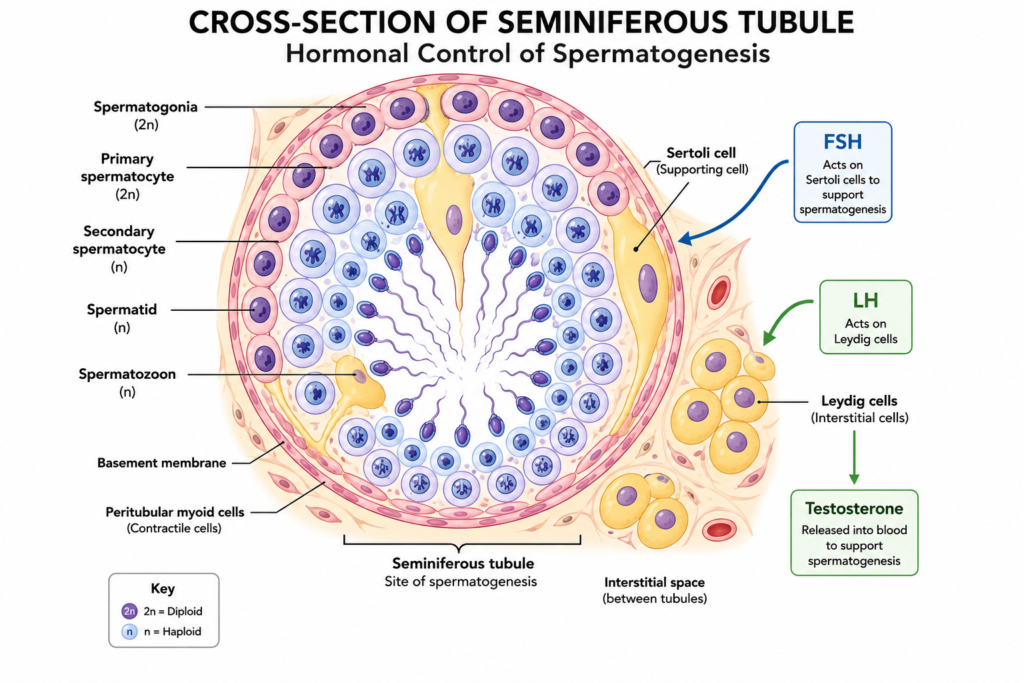
None of this happens automatically — it’s driven by the hypothalamic-pituitary-gonadal (HPG) axis. The hypothalamus releases GnRH (gonadotropin-releasing hormone), which signals the pituitary gland to secrete two hormones: luteinizing hormone (LH) and follicle-stimulating hormone (FSH). These two hormones act on two different cell populations inside the testes. LH acts on Leydig cells, located between the seminiferous tubules, and drives them to produce testosterone. FSH, meanwhile, acts on Sertoli cells, which line the seminiferous tubules and physically support and nourish the developing sperm cells at every stage. Testosterone produced by the Leydig cells then acts on the Sertoli cells as well, and together, testosterone and FSH create the local environment needed for germ cells to survive meiosis and complete their transformation into sperm. Sertoli cells also form the blood-testis barrier, which shields developing sperm from the immune system, since sperm carry unique surface proteins the body would otherwise treat as foreign.
When Spermatogenesis Goes Wrong
Because spermatogenesis is a continuous, high-turnover process, it’s also fairly sensitive to disruption. Conditions like low sperm count (oligospermia), complete absence of sperm in the semen (azoospermia), poor sperm motility (asthenozoospermia), or abnormal sperm shape (teratozoospermia) can all trace back to hormonal imbalances in the HPG axis, genetic conditions like Klinefelter syndrome, varicocele, infections, heat exposure, or lifestyle factors such as smoking and excessive alcohol use. Because sperm are being produced fresh every 64–74 days, many of these issues are at least partially reversible once the underlying cause is addressed — which isn’t true of egg-related fertility problems, for reasons that will become clear in the next section.
Oogenesis: How Egg Cells Are Made
Oogenesis happens in the ovaries, and its timeline is almost the opposite of spermatogenesis in nearly every way.
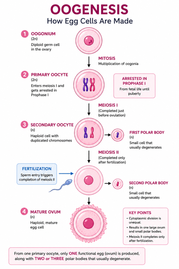
1. Oogonia. These diploid germ cells multiply by mitosis during fetal development. This happens entirely before birth — by the time a baby girl is born, she already has all the egg-precursor cells she will ever have.
2. Primary oocytes. Oogonia develop into primary oocytes and begin meiosis I — but then something unusual happens: the cell arrests, or pauses, in prophase I. This isn’t a brief pause either; primary oocytes stay frozen at this stage for years, sometimes decades, until puberty and then, cycle by cycle, until that particular oocyte is selected for ovulation.
3. Secondary oocyte and first polar body. Once a primary oocyte resumes meiosis I (triggered by the hormonal surge in the menstrual cycle), it divides unequally. Rather than producing two similarly sized cells, it produces one large secondary oocyte and one tiny first polar body. Almost all the cytoplasm and nutrient stores go to the secondary oocyte, since that’s the cell that may eventually support an embryo.
4. Ovulation and the second arrest. The secondary oocyte immediately begins meiosis II — and arrests again, this time in metaphase II. It’s released from the ovary in this paused state during ovulation.
5. Completion — only if fertilized. The secondary oocyte only completes meiosis II if a sperm cell penetrates it. Fertilization triggers the final division, producing a mature ovum and a second polar body. If fertilization doesn’t happen, the secondary oocyte simply degenerates without ever completing meiosis.
The polar bodies produced along the way are essentially “byproducts” — they carry chromosomes but almost no cytoplasm, and they eventually disintegrate. So while spermatogenesis yields four usable sperm, oogenesis yields just one usable egg per meiotic cycle (plus non-functional polar bodies).
Unlike spermatogenesis, oogenesis isn’t continuous. After puberty, generally only one oocyte matures and is released roughly once a month, and this cyclical process continues until menopause, at which point no new oocytes are released.
The Follicle: A Support Structure Sperm Never Needed
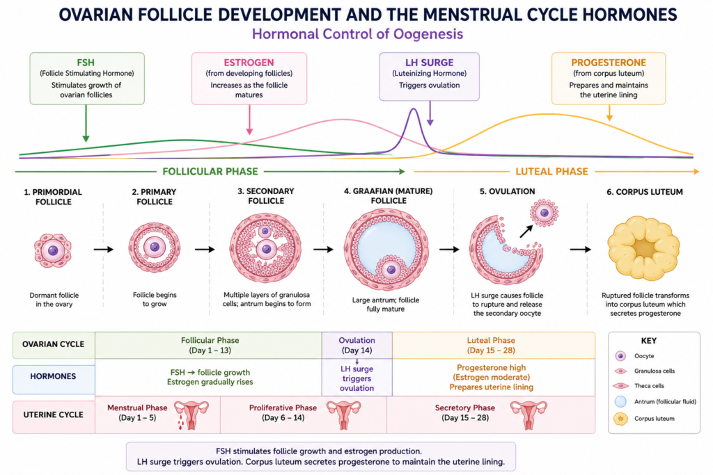
One thing spermatogenesis doesn’t have an equivalent for is the ovarian follicle. Every oocyte develops surrounded by a shell of supporting cells — first granulosa cells alone, and later granulosa cells plus an outer layer of theca cells — and this whole unit, oocyte plus its support cells, is what’s called a follicle. As a follicle matures from a primordial follicle into a fully developed Graafian follicle, it fills with fluid and grows dramatically in size, and the granulosa and theca cells begin actively producing estrogen. Only one follicle typically becomes “dominant” each cycle and goes on to ovulate; the rest degenerate in a process called atresia. This follicular structure is essentially oogenesis’s version of the Sertoli cell support system in the testes — except in females, hundreds of thousands of follicles are recruited and lost over a lifetime for every one that actually completes the journey to ovulation.
Hormonal Control of Oogenesis
Oogenesis is regulated by the same hypothalamic-pituitary-gonadal axis as spermatogenesis, but the way FSH and LH are used is different. During the follicular phase of the menstrual cycle, FSH stimulates a group of follicles to begin maturing, and as they grow, the granulosa cells inside them produce increasing amounts of estrogen. Rising estrogen eventually triggers a sharp spike in LH — the “LH surge” — which is the direct trigger for ovulation: it causes the dominant follicle to rupture and release its secondary oocyte. After ovulation, LH also drives the remnants of the follicle to transform into the corpus luteum, a temporary hormone-producing structure that secretes progesterone to prepare the uterine lining for a possible pregnancy. If fertilization doesn’t occur, the corpus luteum breaks down, hormone levels fall, and menstruation begins — resetting the cycle.
When Oogenesis Goes Wrong
Because oocytes are formed once, before birth, and then sit arrested for years or decades, they’re especially vulnerable to age-related damage — mainly errors in how chromosomes separate during meiosis, a phenomenon called nondisjunction. This is the main reason the risk of chromosomal conditions like Down syndrome rises noticeably as maternal age increases: the longer a primary oocyte has been arrested in prophase I, the more time there’s been for the machinery holding chromosome pairs together to weaken. Other conditions affecting oogenesis include polycystic ovary syndrome (PCOS), where hormonal imbalances disrupt normal follicle maturation and ovulation, premature ovarian insufficiency, where the ovarian reserve is depleted earlier than expected, and Turner syndrome, a chromosomal condition that can cause the ovarian follicle pool to be lost abnormally fast. Unlike male infertility, which often stems from a disruption to an ongoing production line, most female fertility issues relate either to the finite, non-renewable oocyte pool itself or to the hormonal signaling that governs which follicle matures and when.
Difference Between Spermatogenesis and Oogenesis
Here’s a side-by-side comparison of the two processes:
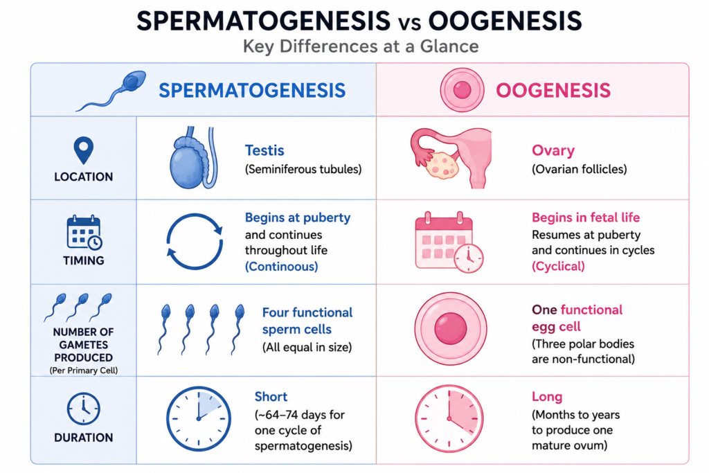
| Feature | Spermatogenesis | Oogenesis |
|---|---|---|
| Location | Seminiferous tubules of the testes | Ovaries |
| Begins | At puberty | Before birth (during fetal development) |
| Continuity | Continuous, from puberty until death | Cyclical (roughly monthly) from puberty until menopause |
| Starting cell | Spermatogonium | Oogonium |
| Cell size through division | Divisions are roughly equal in size | Divisions are unequal — cytoplasm is retained by one cell |
| Number of functional gametes per starting cell | 4 functional sperm | 1 functional egg + 2–3 non-functional polar bodies |
| Meiotic arrest | No arrest — proceeds straight through | Arrests twice: at prophase I (for years) and metaphase II (until fertilization) |
| Completion of meiosis II | Completed independently, before release | Only completed if fertilization occurs |
| Duration | About 64–74 days per cycle | Can take years to decades (from fetal arrest to ovulation) |
| Motility | Sperm are small and motile (flagellum) | Eggs are large, non-motile, and nutrient-rich |
| Output over a lifetime | Hundreds of millions of sperm daily | A finite, pre-set number of oocytes, released one at a time |
| Supporting structure | Sertoli cells within the seminiferous tubules | Granulosa and theca cells forming the ovarian follicle |
| Key hormones involved | FSH (acts on Sertoli cells), LH (drives testosterone via Leydig cells) | FSH (drives follicle growth), LH (triggers ovulation and corpus luteum formation) |
| Common associated disorders | Oligospermia, azoospermia, varicocele, Klinefelter syndrome | PCOS, premature ovarian insufficiency, Turner syndrome, age-related aneuploidy |
| Cell size of gamete | Small, compact, minimal cytoplasm | Large, one of the biggest cells in the human body |
The underlying logic behind these differences comes down to each gamete’s job. Sperm are built for speed, numbers, and mobility — they need to travel and only their DNA needs to make it to the egg. Eggs, on the other hand, are built for provisioning — they need to carry enough cytoplasm, organelles, and nutrients to support the earliest stages of embryonic development, so nature sacrifices quantity for quality by concentrating nearly all the cytoplasm into a single surviving cell.
Why This Comparison Matters
Beyond exams, understanding these two processes has real relevance in reproductive medicine. Age-related fertility decline in women, for instance, is tied directly to the fact that oocytes are formed once, before birth, and simply age along with the rest of the body — increasing the risk of chromosomal errors like nondisjunction the longer a primary oocyte sits arrested in prophase I. Male fertility issues, by contrast, are usually tied to problems arising during the continuous, ongoing production process itself, such as low sperm count or poor motility, rather than the aging of a fixed cell pool.
The Evolutionary Logic Behind the Asymmetry
It’s worth stepping back and asking why these two processes evolved so differently in the first place, since they’re solving what looks like the same problem. The answer usually comes down to a concept evolutionary biologists call anisogamy — the evolution of two distinctly sized gamete types instead of one uniform size. Once one gamete type starts investing more in cytoplasm and nutrients (the “egg strategy”), the other gamete type is free to specialize in numbers and mobility instead (the “sperm strategy”), since it no longer needs to provision the zygote itself. This division of labor is thought to be more efficient overall than both gametes trying to do everything: it guarantees that fertilization produces a zygote with enough resources to begin development (thanks to the egg) while maximizing the odds that fertilization happens at all (thanks to the sheer number and mobility of sperm). This same basic pattern — one large, immobile, resource-rich gamete and one small, mobile, numerous gamete — shows up across most of the animal kingdom, not just in humans, which suggests it’s a deeply conserved evolutionary solution rather than a human-specific quirk.
Frequently Asked Questions
Is gametogenesis the same as meiosis?
Not exactly. Meiosis is the type of cell division that gametogenesis relies on. Gametogenesis is the broader biological process — including cell growth, differentiation, and structural changes — that uses meiosis as one of its steps.
Why does oogenesis produce only one egg but spermatogenesis produces four sperm?
Because oogenesis divides its cytoplasm unequally to concentrate resources into a single large, viable egg, while spermatogenesis divides its cytoplasm equally since sperm don’t need to carry nutritional reserves.
Why do primary oocytes stay arrested for so long?
The arrest allows oocytes to remain in a stable, dormant state until hormonal signals during each menstrual cycle trigger further development — this is part of why oocyte quality can decline with maternal age.
What triggers ovulation?
A sharp surge in luteinizing hormone (LH), caused by rising estrogen from the maturing dominant follicle, is the direct trigger that causes the follicle to rupture and release the secondary oocyte.
Why is male fertility often more treatable than female fertility issues?
Because spermatogenesis is an ongoing production process — a fresh batch of sperm is generated roughly every two to two-and-a-half months — many causes of low sperm quality can improve once the underlying issue (hormonal, lifestyle, or structural) is corrected. Oogenesis, by contrast, works with a fixed, non-renewable pool of oocytes established before birth, so age-related decline in egg quality or quantity generally can’t be reversed.
Do Sertoli cells and granulosa cells serve the same purpose?
Functionally, yes — both are somatic support cells that nurture the developing germ cell, respond to pituitary hormones, and create the local hormonal environment needed for gamete maturation, even though they sit in anatomically different organs.
References
- Gilbert, S.F. — Spermatogenesis, Developmental Biology, 6th Edition — NCBI Bookshelf
- McCarrey, J.R. — Spermatogenesis–oogenesis: a cyto-morphological comparison — PubMed, Biology of Reproduction
- Sanford, J.P. et al. — Differences in DNA methylation during oogenesis and spermatogenesis — PubMed, Genes & Development
- 43.3C: Gametogenesis (Spermatogenesis and Oogenesis) — Biology LibreTexts
- Spermatogenesis vs. Oogenesis: Differences and Examples — Microbe Notes
- Role of Follicle-Stimulating Hormone in Spermatogenesis — Frontiers in Endocrinology
- The Role of Luteinizing Hormone Activity in Spermatogenesis — Reproductive Biology and Endocrinology, Springer Nature
- Physiology, Menstrual Cycle — StatPearls, NCBI Bookshelf, NIH
- 16.6 Female Puberty, Ovarian Cycle, and Menstrual Cycle — General Anatomy & Physiology, Pressbooks
Discover more from Zoologyverse
Subscribe to get the latest posts sent to your email.