
Lymphoid organs are organized structures of the immune system in which lymphocytes develop, mature, circulate, encounter antigens, and participate in immune responses. They provide specialized environments where immune cells interact with one another and with supporting cells.
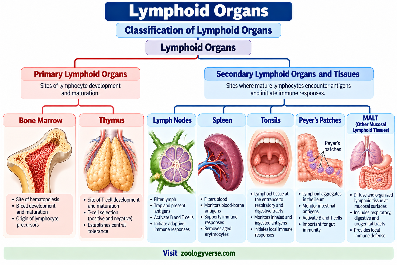
The major lymphoid organs are traditionally divided into primary lymphoid organs and secondary lymphoid organs. The primary lymphoid organs are the bone marrow and thymus, where lymphocytes develop and mature. The major secondary lymphoid organs and tissues include lymph nodes, spleen, tonsils, Peyer’s patches, and other mucosa-associated lymphoid tissue (MALT).
The organization of these structures is essential for both innate and adaptive immune functions. Primary lymphoid organs provide suitable environments for lymphocyte development, whereas secondary lymphoid organs bring mature lymphocytes into contact with antigens and antigen-presenting cells.
Understanding lymphoid organs is therefore important for studying the immune system, lymphocyte development, antigen recognition, antibody production, and adaptive immune responses.
This article explains the major lymphoid organs, their classification, structure, functions, and importance in immunology.
What Are Lymphoid Organs?
Lymphoid organs are organized lymphoid tissues containing large numbers of lymphocytes supported by a framework of non-lymphoid cells and extracellular structures.
Their major functions include:
- Development and maturation of lymphocytes.
- Maintenance of mature lymphocytes.
- Transport and concentration of antigens.
- Interaction between lymphocytes and antigen-presenting cells.
- Initiation of adaptive immune responses.
- Production of antibodies following appropriate immune activation.
- Formation and maintenance of immunological memory.
The organization of lymphoid organs allows immune cells to move between blood, lymphatic vessels, tissues, and specialized immune compartments.
The two major functional categories are primary lymphoid organs and secondary lymphoid organs.
Classification of Lymphoid Organs
The lymphoid organs can be broadly classified into two major groups.
| Type | Major Lymphoid Organs | Main Function |
|---|---|---|
| Primary lymphoid organs | Bone marrow and thymus | Development and maturation of lymphocytes |
| Secondary lymphoid organs and tissues | Lymph nodes, spleen, tonsils, Peyer’s patches and MALT | Antigen encounter, lymphocyte activation and adaptive immune responses |
Primary Lymphoid Organs
The primary lymphoid organs are the bone marrow and thymus in humans and other mammals.
Lymphocyte precursors originate from hematopoietic stem cells in the bone marrow. B lymphocytes undergo their major maturation process in the bone marrow, whereas T-cell precursors migrate to the thymus for T-cell maturation and selection.
Secondary Lymphoid Organs
Secondary lymphoid organs and tissues are sites where mature lymphocytes encounter antigens and where many adaptive immune responses are initiated.
Important secondary sites include:
- Lymph nodes
- Spleen
- Tonsils
- Peyer’s patches
- Mucosa-associated lymphoid tissue
- Other organized lymphoid tissues associated with mucosal surfaces
The lymph nodes primarily monitor antigens arriving from tissues through lymph, while the spleen monitors antigens present in the blood. MALT is positioned at mucosal surfaces where environmental antigens and microorganisms may enter the body.
Bone Marrow
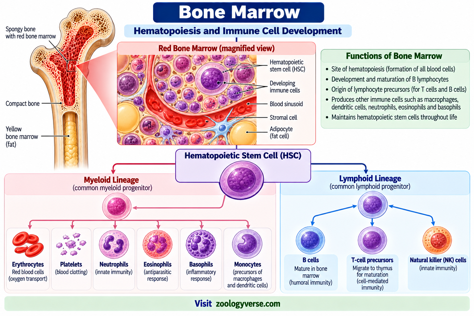
The bone marrow is a primary lymphoid organ and an important site of hematopoiesis. Hematopoietic stem cells in the bone marrow give rise to the different blood-cell lineages, including cells that participate in immune defense.
In mammals, B-cell development and maturation occur in the bone marrow, while T-cell precursors leave the bone marrow and migrate to the thymus.
Functions of Bone Marrow
The bone marrow performs several important functions:
- Produces blood cells through hematopoiesis.
- Generates lymphocyte precursors.
- Provides the developmental environment for B lymphocytes.
- Produces new immune cells throughout life.
- Provides a reservoir and maintenance environment for several mature immune-cell populations.
In adults, active red bone marrow is concentrated mainly in bones such as the pelvis, vertebrae, ribs, sternum, and other axial skeletal regions.
Bone marrow is therefore much more than a simple blood-cell-producing tissue. It provides an essential developmental environment for the immune system.
Bone Marrow and B-Cell Development
Both B and T lymphocyte precursors originate from hematopoietic stem cells in the bone marrow. B lymphocytes continue their major development and maturation there, whereas T-cell precursors migrate to the thymus.
After maturation, naïve B and T lymphocytes enter the circulation and travel through secondary lymphoid organs, where they can encounter their specific antigens.
Thymus
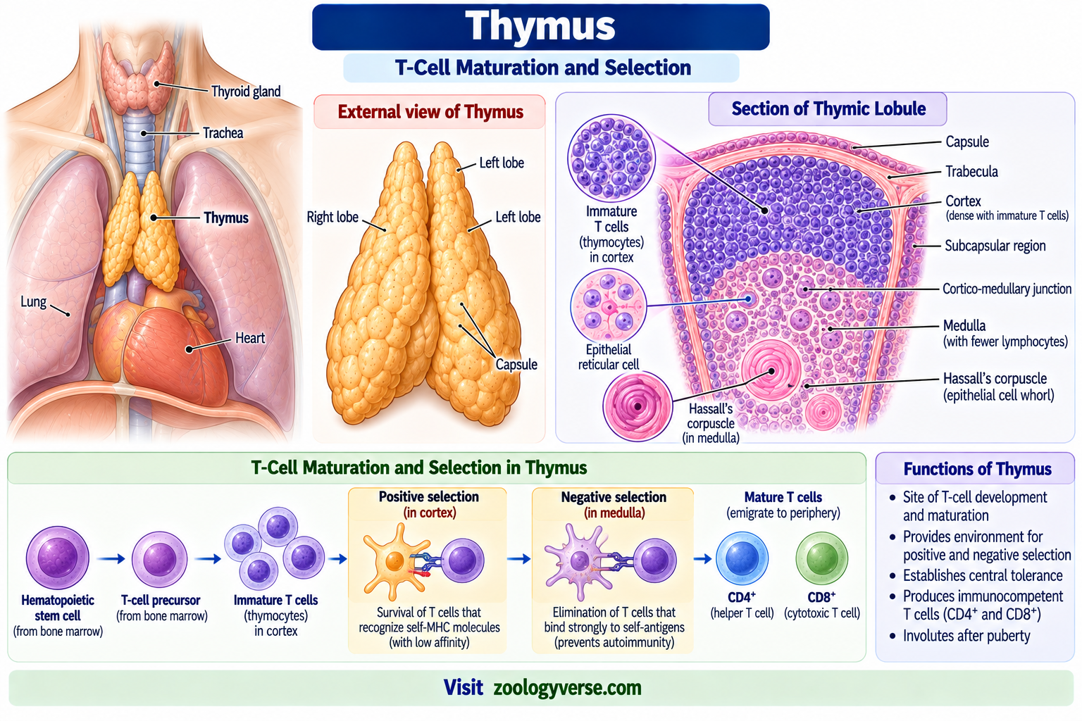
The thymus is a primary lymphoid organ specialized for T-lymphocyte development and selection.
It is located in the upper anterior chest, behind the sternum. The thymus is relatively large and active during childhood and undergoes progressive involution after puberty, although thymic function does not necessarily disappear completely in adulthood.
The thymus provides the specialized microenvironment required for developing T cells to undergo differentiation and selection.
Structure of the Thymus
The thymus is divided into lobules containing two major regions:
- Cortex
- Medulla
The cortex contains a high density of developing T lymphocytes, known as thymocytes.
The medulla contains fewer developing lymphocytes and contains specialized epithelial cells, dendritic cells, macrophages, and characteristic Hassall’s corpuscles.
Functions of the Thymus
The major functions of the thymus include:
- T-lymphocyte development.
- T-cell receptor formation and maturation.
- Positive selection of developing T cells.
- Negative selection of strongly self-reactive T cells.
- Establishment of an appropriate functional T-cell repertoire.
- Contribution to central immune tolerance.
T-cell selection is essential because mature T cells must be capable of recognizing antigen presented by self-MHC molecules while avoiding strong reactivity against the body’s own tissues.
The thymus is therefore one of the most important primary lymphoid organs for adaptive immunity.
Lymph Nodes
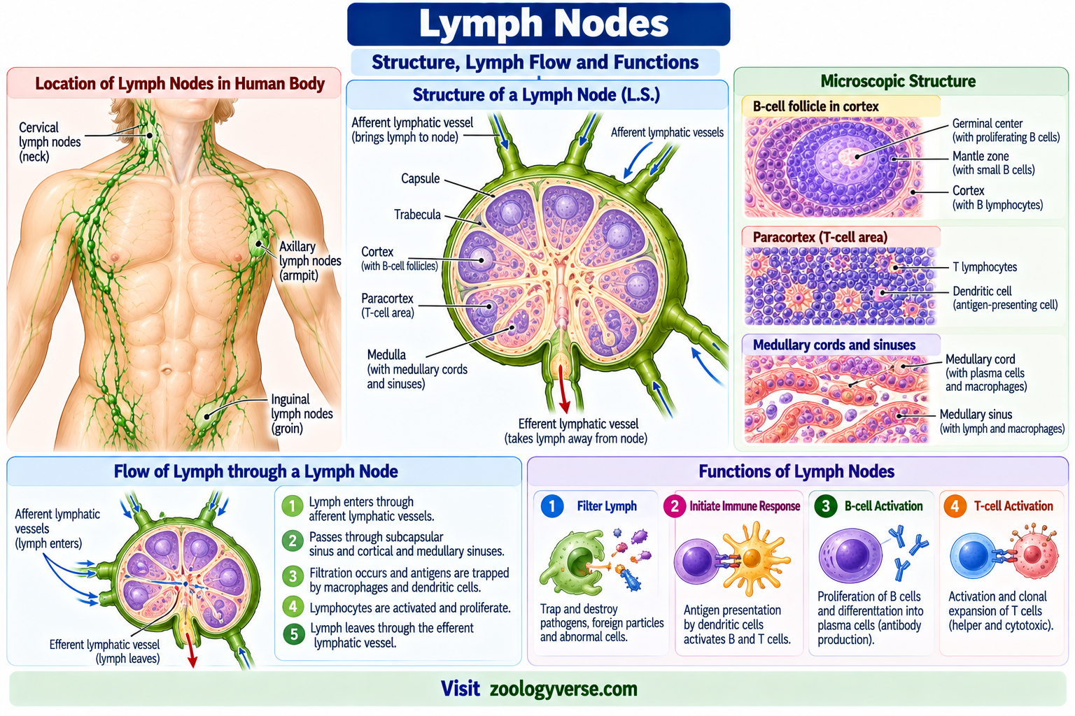
Lymph nodes are small, organized secondary lymphoid organs located along lymphatic vessels.
They act as surveillance and immune-response sites for antigens and antigen-presenting cells arriving from tissues through lymphatic vessels.
A lymph node contains specialized regions that organize B cells, T cells, antigen-presenting cells, and other immune cells.
Structure of a Lymph Node
A typical lymph node has:
- Fibrous capsule
- Subcapsular sinus
- Cortex
- Paracortex
- Medulla
- Afferent lymphatic vessels
- Efferent lymphatic vessel
- Blood vessels
The cortex contains lymphoid follicles that are rich in B lymphocytes.
The paracortex is relatively rich in T lymphocytes and contains important antigen-presenting cells.
The medulla contains medullary cords and sinuses through which lymph and immune cells pass.
Functions of Lymph Nodes
The major functions of lymph nodes include:
- Filtering lymph.
- Trapping and concentrating antigens.
- Providing sites for antigen presentation.
- Supporting interaction between dendritic cells and T cells.
- Supporting B-cell activation.
- Supporting antibody responses.
- Allowing proliferation and differentiation of activated lymphocytes.
When an infection occurs in a tissue, antigen-bearing dendritic cells and other antigenic material can travel through lymphatic vessels to the draining lymph node.
There, appropriate lymphocytes can become activated and undergo clonal expansion and differentiation.
B-Cell Follicles and Germinal Centers
B cells are concentrated in lymphoid follicles within lymph nodes.
Following appropriate antigen stimulation, activated B cells can form germinal centers where they undergo proliferation and further differentiation with the help of specialized cells and signals.
These processes can ultimately generate antibody-secreting plasma cells and memory B cells.
Spleen
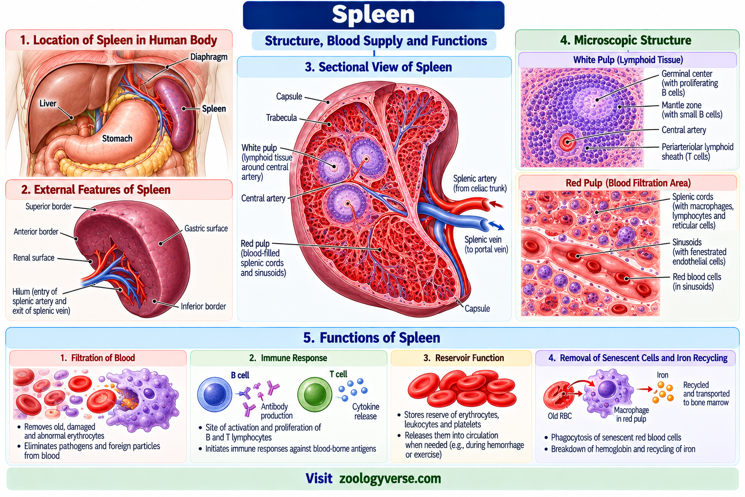
The spleen is a major secondary lymphoid organ that monitors the blood.
Unlike lymph nodes, which primarily receive lymph draining from tissues, the spleen is specialized for immune surveillance of blood-borne antigens.
The spleen is located in the upper left region of the abdomen and has important immune and hematological functions.
Structure of the Spleen
The spleen contains two major functional regions:
- White pulp
- Red pulp
The white pulp contains lymphoid tissue involved in immune responses to blood-borne antigens.
The red pulp contains vascular spaces and cords involved in blood filtration and removal of aged or damaged red blood cells.
White Pulp
White pulp contains organized lymphoid areas associated with the splenic circulation.
It includes regions enriched in B cells and T cells and provides an environment in which immune cells can respond to antigens arriving in the blood.
Red Pulp
The red pulp is involved mainly in blood filtration.
Important functions include:
- Removal of aged or damaged erythrocytes.
- Removal of some cellular debris.
- Blood storage in some species.
- Participation in immune surveillance of blood.
Functions of the Spleen
The spleen:
- Monitors blood-borne antigens.
- Supports adaptive immune responses.
- Contains B and T lymphocytes.
- Supports antigen presentation.
- Removes aged and damaged erythrocytes.
- Participates in clearance of certain blood-borne particles and microorganisms.
The spleen is therefore an important component of the network of secondary lymphoid organs.
Mucosa-Associated Lymphoid Tissue (MALT)
Mucosa-associated lymphoid tissue (MALT) refers to organized and diffuse lymphoid tissue associated with mucosal surfaces.
Mucosal surfaces are continuously exposed to food molecules, environmental substances and microorganisms. MALT provides immune surveillance at these important interfaces.
MALT occurs in several locations, particularly in the gastrointestinal and respiratory tracts.
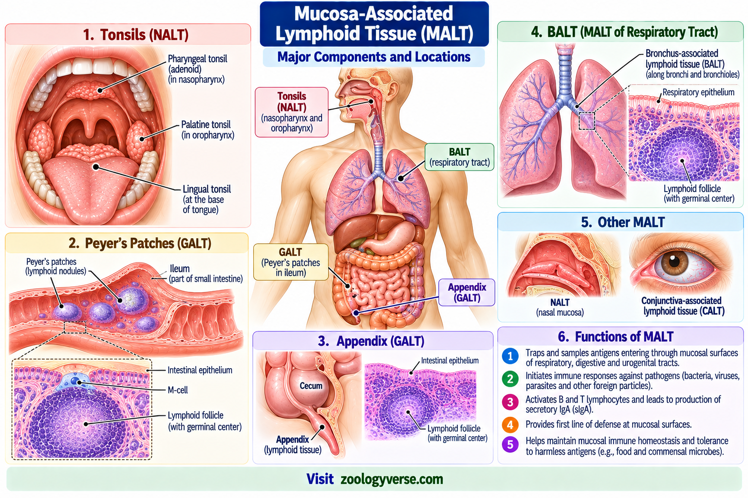
Examples include:
- Tonsils
- Peyer’s patches
- Other intestinal lymphoid aggregates
- Bronchus-associated lymphoid tissue in appropriate contexts
- Other mucosal lymphoid tissues
MALT can contain B cells, T cells, antigen-presenting cells, macrophages and other immune cells.
Functions of MALT
The major functions of MALT include:
- Sampling and monitoring mucosal antigens.
- Supporting local immune responses.
- Activating B and T lymphocytes.
- Supporting antibody responses at mucosal surfaces.
- Contributing to protection against invading microorganisms.
MALT is particularly important because many pathogens enter the body through mucosal surfaces.
Tonsils
Tonsils are secondary lymphoid tissues associated with the entrance to the respiratory and digestive tracts.
Major groups include:
- Palatine tonsils
- Pharyngeal tonsil
- Lingual tonsils
They contain lymphoid follicles and immune cells that participate in surveillance of material entering through the mouth and nose.
Functions of Tonsils
Tonsils:
- Monitor inhaled and ingested materials.
- Contain lymphoid follicles.
- Support local immune responses.
- Participate in antigen recognition at mucosal surfaces.
Tonsils therefore represent an important component of mucosal immune defense.
Peyer’s Patches
Peyer’s patches are organized lymphoid aggregates located mainly in the ileum of the small intestine.
They are an important component of intestinal mucosal immunity.
Peyer’s patches contain B cells, T cells, antigen-presenting cells and specialized epithelial structures that facilitate antigen sampling.
Functions of Peyer’s Patches
Their functions include:
- Sampling intestinal antigens.
- Supporting mucosal immune responses.
- Activating lymphocytes.
- Supporting B-cell responses.
- Contributing to immune surveillance of the intestinal tract.
Peyer’s patches are therefore an important example of organized mucosal lymphoid tissue.
Primary vs Secondary Lymphoid Organs
The distinction between primary and secondary lymphoid organs is based mainly on their roles in lymphocyte development versus immune activation.
| Feature | Primary Lymphoid Organs | Secondary Lymphoid Organs |
|---|---|---|
| Major examples | Bone marrow and thymus | Lymph nodes, spleen, tonsils and MALT |
| Main role | Lymphocyte development and maturation | Antigen encounter and immune activation |
| B-cell maturation | Bone marrow | Activated B cells respond and differentiate |
| T-cell maturation | Thymus | Mature T cells encounter antigen |
| Antigen-driven adaptive response | Not the main function | Major function |
| Major cell interactions | Developing lymphocytes and stromal cells | Mature lymphocytes, APCs and antigen |
| Examples of immune response | Selection and maturation | Clonal expansion and effector responses |
The primary and secondary lymphoid organs are functionally connected. Mature lymphocytes leave the primary organs and continuously circulate through blood and secondary lymphoid tissues.
Structure and Functions of Lymphoid Organs
Although different lymphoid organs have different structures, they share a common principle: specialized organization allows immune cells to encounter antigens and communicate efficiently.
For example:
- Bone marrow provides a developmental environment.
- Thymus supports T-cell selection.
- Lymph nodes concentrate antigens from tissues.
- Spleen monitors blood-borne antigens.
- MALT monitors mucosal surfaces.
This functional specialization allows the immune system to detect threats at different anatomical locations.
Role of Lymphoid Organs in Immune Responses
The lymphoid organs are essential for coordinating immune responses.
A simplified sequence can be described as:
Pathogen enters tissue
↓
Innate immune recognition
↓
Antigen capture by dendritic cells and other APCs
↓
Transport of antigen or antigen-bearing cells to a secondary lymphoid organ
↓
Activation of antigen-specific lymphocytes
↓
Clonal expansion and differentiation
↓
Effector immune response
↓
Memory-cell formation
For example, dendritic cells can carry antigen from an infected tissue to a draining lymph node. There, they can present antigen-derived peptides to naïve T cells and help initiate an adaptive immune response.
B cells can also become activated in secondary lymphoid tissues and eventually differentiate into plasma cells that produce antibodies.
These processes demonstrate why secondary lymphoid organs are central to adaptive immunity.
Lymphoid Organs vs Lymphoid Tissues
The terms lymphoid organ and lymphoid tissue are closely related but are not always used identically in different textbooks.
Lymphoid organs are highly organized structures with defined anatomical architecture, such as lymph nodes, spleen, thymus and bone marrow.
Lymphoid tissues can also occur as diffuse or organized aggregates associated with mucosal surfaces.
MALT is therefore often discussed as a group of secondary lymphoid tissues rather than as a single anatomical organ.
For examination purposes, the important distinction is that both organized lymphoid organs and lymphoid tissues provide specialized environments for immune-cell development, surveillance or immune responses.
How Lymphoid Organs Connect With the Cells of the Immune System
The function of lymphoid organs cannot be understood without considering the immune cells that occupy them.
B cells mature mainly in the bone marrow and later populate secondary lymphoid tissues.
T cells mature in the thymus before entering the circulation and secondary lymphoid organs.
Dendritic cells capture antigens in tissues and can transport antigen-derived information to lymph nodes.
Macrophages contribute to antigen processing, phagocytosis and tissue immune defense.
Plasma cells can develop from activated B cells and produce antibodies.
The relationship between these cells and their anatomical environments is fundamental to effective immunity.
For a detailed discussion of these immune cells, see the internal article Cells of the Immune System.
Lymphoid Organs and Innate Immunity
Although lymphoid organs are strongly associated with adaptive immunity, they also contain many cells involved in innate immune defense.
Macrophages, dendritic cells, natural killer cells and other innate immune cells are present in lymphoid tissues and participate in early immune responses.
Innate immune signals can influence the activation and differentiation of adaptive lymphocytes.
This illustrates the close relationship between innate immunity and adaptive immunity.
For an overview of this relationship, see Innate Immunity vs Adaptive Immunity.
Lymphoid Organs and Adaptive Immunity
Secondary lymphoid organs are especially important for adaptive immunity because they provide locations where mature lymphocytes encounter antigen.
Naïve T cells continuously recirculate through secondary lymphoid tissues. When a T cell encounters an appropriate antigen-MHC complex presented by an antigen-presenting cell, it can become activated and proliferate.
B cells can similarly encounter antigen and receive the signals necessary for activation and differentiation.
The resulting effector cells participate in pathogen elimination, while some activated lymphocytes become memory cells.
The broader concept of immune protection can be reviewed in Types of Immunity, which explains innate, adaptive, active and passive forms of immunity.
Key Differences Between Lymphoid Organs
| Lymphoid organ | Type | Major immune function |
|---|---|---|
| Bone marrow | Primary | Hematopoiesis and B-cell development |
| Thymus | Primary | T-cell maturation and selection |
| Lymph nodes | Secondary | Immune responses to tissue-derived antigens |
| Spleen | Secondary | Immune responses to blood-borne antigens |
| Tonsils | Secondary lymphoid tissue | Mucosal immune surveillance |
| Peyer’s patches | Secondary lymphoid tissue | Intestinal immune surveillance |
| MALT | Secondary lymphoid tissue | Mucosal immune defense |
Importance of Lymphoid Organs
The importance of lymphoid organs can be summarized through their major functions:
- Lymphocyte development
Primary lymphoid organs provide specialized environments for lymphocyte development and maturation. - Antigen surveillance
Secondary lymphoid organs collect or encounter antigens from tissues, blood or mucosal surfaces. - Lymphocyte activation
Mature B and T cells can encounter antigen in secondary lymphoid tissues. - Antibody responses
Activated B cells can differentiate into antibody-secreting plasma cells. - Cell-mediated immunity
Activated T cells can coordinate immune responses or eliminate appropriate target cells. - Immune memory
Adaptive immune responses can generate memory B and T cells that contribute to faster responses upon later exposure. - Immune regulation
Specialized cellular environments help regulate the magnitude and type of immune responses.
Frequently Asked Questions
What are lymphoid organs?
What are the primary lymphoid organs?
What are secondary lymphoid organs?
What is the function of lymph nodes?
What is the function of the spleen?
Why is the thymus important?
Is bone marrow a lymphoid organ?
What is MALT?
Where do adaptive immune responses begin?
Key Takeaways
- Lymphoid organs are organized immune tissues containing large populations of lymphocytes.
- They are broadly classified into primary and secondary lymphoid organs.
- The major primary lymphoid organs in mammals are bone marrow and thymus.
- B lymphocytes undergo their major maturation process in the bone marrow.
- T-cell precursors migrate from bone marrow to the thymus for maturation and selection.
- The thymus has a cortex and medulla.
- Hassall’s corpuscles are characteristic structures of the thymic medulla.
- Lymph nodes are secondary lymphoid organs that monitor antigens arriving from tissues through lymph.
- B cells are concentrated mainly in lymphoid follicles of lymph nodes.
- T cells are abundant in the paracortical region.
- The spleen monitors blood-borne antigens.
- The spleen contains white pulp and red pulp.
- MALT is associated with mucosal surfaces.
- Tonsils are important lymphoid tissues at the entrance to the respiratory and digestive tracts.
- Peyer’s patches are organized lymphoid aggregates mainly found in the ileum.
- Secondary lymphoid organs are major sites for antigen-driven adaptive immune responses.
- Mature lymphocytes continuously recirculate between blood and secondary lymphoid tissues.
- Lymphoid organs provide specialized environments that allow immune cells to communicate efficiently.
- Primary organs are mainly associated with lymphocyte development and maturation, whereas secondary organs are mainly associated with antigen encounter and immune activation.
Conclusion
Lymphoid organs form an organized anatomical network that supports the development, maturation, surveillance and activation of immune cells.
The bone marrow and thymus serve as the major primary lymphoid organs in mammals. The bone marrow supports hematopoiesis and B-cell development, while the thymus provides the environment required for T-cell maturation and selection.
The major secondary lymphoid organs, including lymph nodes and spleen, together with mucosal lymphoid tissues such as tonsils and Peyer’s patches, provide sites where mature lymphocytes encounter antigens and initiate adaptive immune responses.
Understanding the structure and functions of lymphoid organs is essential for studying lymphocytes, antigen presentation, antibody responses, cell-mediated immunity and the overall organization of the immune system.
References
- NCBI Bookshelf. Immunobiology: The Components of the Immune System — Lymphocytes Mature in the Bone Marrow or the Thymus.
- NCBI Bookshelf. Generation of Lymphocytes in Bone Marrow and Thymus.
- NCBI Bookshelf. In brief: What are the organs of the immune system?
- NCBI/PMC. Bone Marrow, Blood Cells, and the Lymphoid/Lymphatic System.
- NCBI Bookshelf. Physiology, Immune Response.
Discover more from Zoologyverse
Subscribe to get the latest posts sent to your email.