
The growth, development, repair, and reproduction of living organisms depend on the ability of cells to divide in a controlled manner.
Before a cell divides, it must grow, duplicate its genetic material, and prepare the cellular components required to produce new cells.
The complete sequence of events through which a cell grows and divides is known as the cell cycle.
Cell division occurs mainly through two processes: mitosis and meiosis.
Mitosis produces genetically similar daughter cells and is important for growth, tissue repair, and asexual reproduction.
Meiosis, in contrast, produces genetically different haploid cells and is essential for sexual reproduction and the maintenance of chromosome number across generations.
Understanding the cell cycle and these two forms of cell division is fundamental to cell biology, genetics, development, reproduction, and evolution.
What Is the Cell Cycle?
The cell cycle is the ordered series of events through which a cell grows, duplicates its DNA, and divides into daughter cells.
The cell cycle is broadly divided into two major phases:
- Interphase
- M phase (mitotic or meiotic phase)
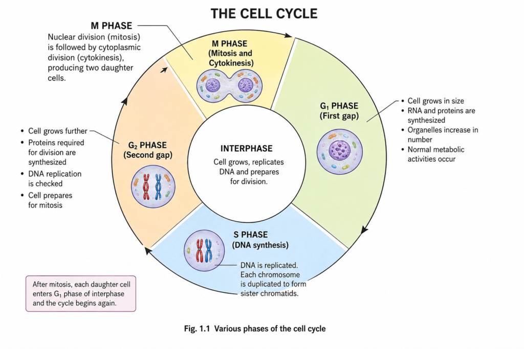
Interphase is further divided into G₁, S, and G₂ phases.
1. G₁ Phase – First Gap Phase
The G₁ phase begins immediately after cell division. During this stage:
- The cell increases in size.
- RNA and proteins are synthesized.
- Cell organelles increase in number.
- Normal cellular activities continue.
- The cell prepares for DNA replication.
A major G₁ checkpoint determines whether the cell has appropriate conditions and sufficient resources to proceed toward DNA replication.
2. S Phase – DNA Synthesis
During the S phase, the cell replicates its DNA.
Each chromosome is duplicated to form two genetically identical sister chromatids joined at the centromere. Although the amount of DNA doubles, the chromosome number does not change.
For example, a diploid cell with 2n chromosomes remains 2n chromosomes, but each chromosome consists of two sister chromatids after DNA replication.
3. G₂ Phase – Second Gap Phase
During G₂:
- The cell continues to grow.
- Proteins required for cell division are synthesized.
- DNA replication is checked.
- The cell prepares for chromosome segregation.
The G₂ checkpoint helps ensure that DNA replication has been completed correctly before the cell enters division.
4. M Phase – Cell Division
The M phase involves nuclear division and usually cytokinesis.
Depending on the biological context, nuclear division can occur through:
- Mitosis, producing two genetically similar daughter cells.
- Meiosis, producing haploid cells involved in sexual reproduction.
Cell Cycle Checkpoints
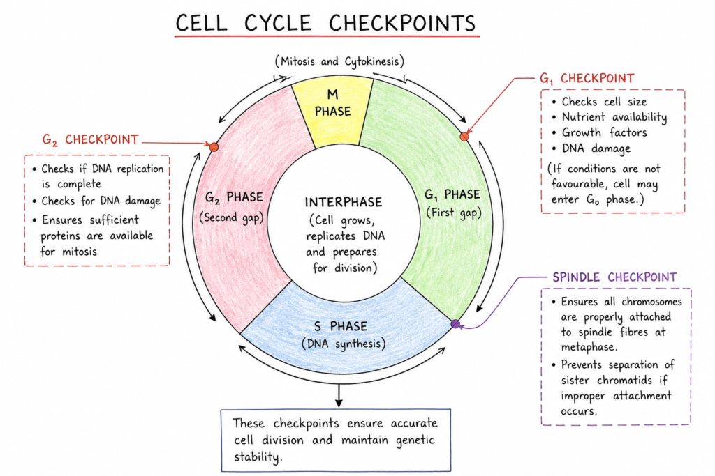
Cell-cycle progression is tightly regulated by checkpoints that help prevent cells with damaged or incompletely replicated DNA from dividing.
The major checkpoints include:
- G₁ checkpoint: Determines whether the cell is ready to replicate its DNA.
- G₂ checkpoint: Checks whether DNA replication has been completed properly.
- Spindle checkpoint: Ensures that chromosomes are correctly attached to spindle fibres before separation.
Regulation of the cell cycle involves proteins such as cyclins and cyclin-dependent kinases (CDKs). Failure of these regulatory mechanisms can contribute to uncontrolled cell proliferation and cancer.
Mitotic Cell Division
Mitosis is a type of nuclear division in which duplicated chromosomes are separated so that each daughter cell receives an essentially identical set of chromosomes.
Mitosis is generally followed by cytokinesis, which divides the cytoplasm.
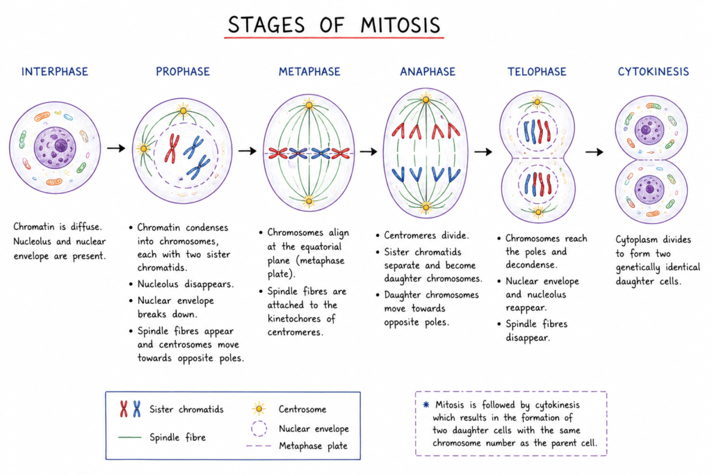
Mitosis is traditionally divided into four major stages:
- Prophase
- Metaphase
- Anaphase
- Telophase
Cytokinesis usually follows or overlaps with telophase.
1. Prophase
During prophase:
- Chromatin condenses into visible chromosomes.
- Each chromosome consists of two sister chromatids.
- The mitotic spindle begins to form.
- The nucleolus disappears.
- The centrosomes move toward opposite poles in animal cells.
In many textbook descriptions, the breakdown of the nuclear envelope is associated with prometaphase, which follows prophase.
2. Metaphase
During metaphase:
- Chromosomes become highly condensed.
- Spindle microtubules attach to chromosome kinetochores.
- Chromosomes align near the equatorial plane of the cell.
This arrangement helps ensure accurate chromosome segregation.
3. Anaphase
During anaphase:
- Sister chromatids separate.
- Each separated chromatid becomes an individual daughter chromosome.
- Spindle fibres move the chromosomes toward opposite poles.
This ensures that each future daughter cell receives one copy of each chromosome.
4. Telophase
During telophase:
- Chromosomes reach opposite poles.
- Chromosomes begin to decondense.
- Nuclear envelopes form around the separated chromosome sets.
- Nucleoli reappear.
5. Cytokinesis
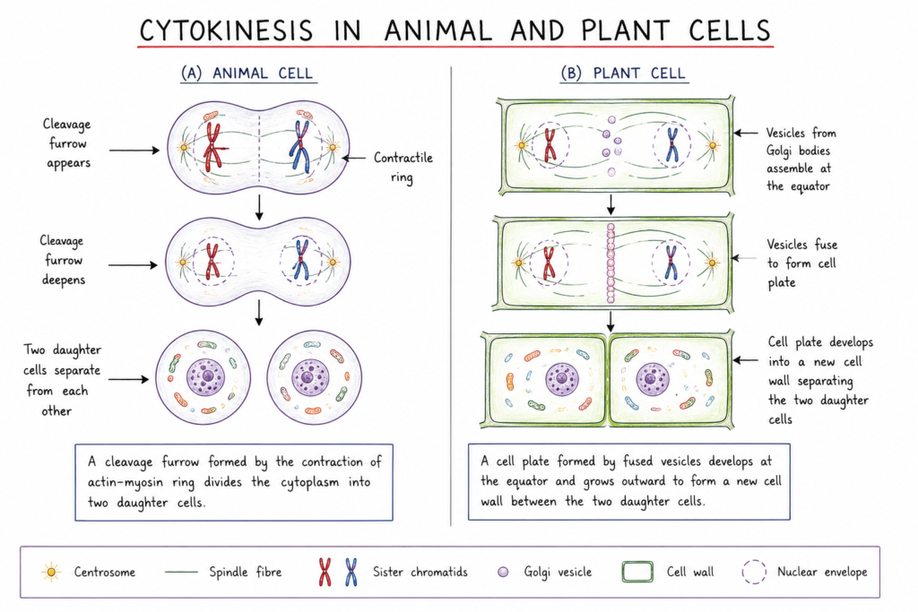
Cytokinesis divides the cytoplasm into two daughter cells.
In animal cells, a cleavage furrow constricts the cell membrane.
In plant cells, vesicles accumulate at the equatorial region and form a cell plate, which develops into a new cell wall between the daughter cells.
Significance of Mitosis
Mitosis is essential for multicellular organisms because it maintains chromosome number and produces cells required for growth and maintenance.
Its major functions include:
Growth and Development
Repeated mitotic divisions increase the number of cells during embryonic and post-embryonic development.
Tissue Repair
Mitosis replaces cells damaged through injury or normal wear and tear.
Cell Replacement
Many tissues continuously replace old or damaged cells through mitotic division.
Asexual Reproduction
Mitosis contributes to asexual reproduction in many organisms, including some unicellular organisms and multicellular organisms that reproduce vegetatively.
Genetic Stability
Because daughter cells normally receive equivalent chromosome sets, mitosis helps maintain genetic stability from one somatic cell generation to the next.
Meiotic Cell Division
Meiosis is a specialized form of cell division associated with sexual reproduction. It involves one round of DNA replication followed by two successive nuclear divisions, called meiosis I and meiosis II.
Meiosis ultimately produces haploid cells from a diploid starting cell.
A crucial feature of meiosis is that it generates genetic variation through:
- Crossing over
- Independent assortment of homologous chromosomes
- Random combination of parental chromosomes
Stages of Meiosis
Meiosis consists of two divisions:
DNA replication occurs before meiosis I, during the S phase. There is no second round of DNA replication between meiosis I and meiosis II.
Meiosis I
1. Prophase I
Prophase I is the longest and most complex stage of meiosis.
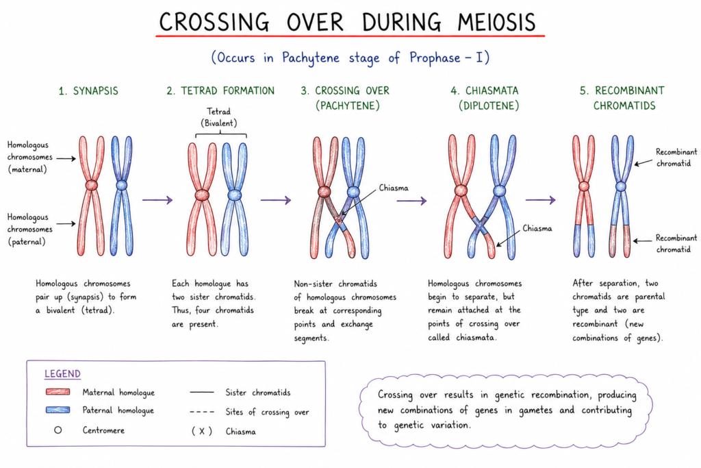
Homologous chromosomes pair with one another in a process called synapsis, forming structures known as bivalents or tetrads.
Prophase I is traditionally divided into five substages:
- Leptotene
- Zygotene
- Pachytene
- Diplotene
- Diakinesis
Crossing Over
During pachytene, homologous chromosomes exchange corresponding segments of DNA through a process called crossing over.
The visible points where homologous chromosomes remain associated after crossing over are called chiasmata.
Crossing over creates new combinations of alleles and is an important source of genetic variation.
2. Metaphase I
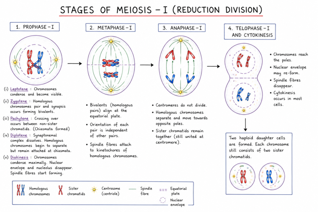
Homologous chromosome pairs align at the equatorial region of the cell.
The orientation of each homologous pair is independent of other chromosome pairs. This contributes to independent assortment.
3. Anaphase I
Homologous chromosomes separate and move toward opposite poles.
Importantly, sister chromatids remain together during anaphase I.
This is the key event responsible for reducing the chromosome number from diploid to haploid.
4. Telophase I and Cytokinesis
Chromosomes reach opposite poles and cytokinesis may occur.
Depending on the organism, nuclear envelopes may reform temporarily.
The resulting cells are haploid, but each chromosome still consists of two sister chromatids.
Meiosis II
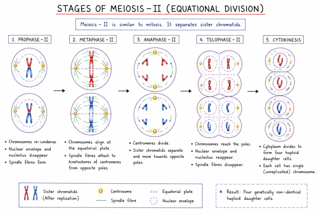
Meiosis II resembles mitosis because sister chromatids are separated.
1. Prophase II
Chromosomes condense again and a new spindle apparatus forms.
2. Metaphase II
Chromosomes align individually at the equatorial plane.
3. Anaphase II
The centromeres separate, allowing sister chromatids to move toward opposite poles.
4. Telophase II and Cytokinesis
Nuclei reform around the chromosome sets, chromosomes decondense, and cytokinesis occurs.
The final result is generally four haploid cells, although the exact products differ between organisms and between male and female gametogenesis.
Significance of Meiosis
Meiosis has several important biological roles.
Maintenance of Chromosome Number
Meiosis reduces the chromosome number by half, producing haploid gametes in organisms where gametes are the products of meiosis.
During fertilization, fusion of haploid gametes restores the diploid chromosome number.
Genetic Variation
Meiosis is one of the major sources of genetic variation.
Variation results from:
- Crossing over
- Independent assortment
- Random segregation of homologous chromosomes
This variation provides the raw material on which natural selection can act.
Sexual Reproduction
Meiosis is essential for producing reproductive cells in animals and for generating spores in many plants and fungi.
Evolutionary Significance
By generating genetically diverse reproductive cells, meiosis contributes to variation within populations and therefore plays an important role in evolutionary processes.
Mitosis vs Meiosis
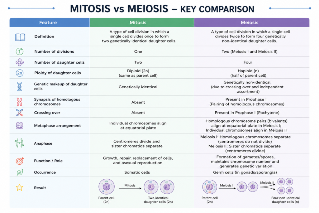
| Feature | Mitosis | Meiosis |
|---|---|---|
| Number of divisions | One | Two |
| DNA replication | Once before division | Once before meiosis I |
| Daughter cells | Usually two | Usually four |
| Chromosome number | Maintained | Reduced by half |
| Genetic similarity | Usually genetically similar to parent cell | Genetically varied |
| Homologous chromosome pairing | Absent | Present in prophase I |
| Crossing over | Normally absent | Present during prophase I |
| Main role | Growth, repair, cell replacement | Sexual reproduction |
| Separation in first division | Sister chromatids | Homologous chromosomes |
| Genetic variation | Limited | High |
Mitosis and Meiosis: Biological Importance
Mitosis and meiosis perform different but complementary roles.
Mitosis maintains the organism. It increases cell number, replaces damaged cells, and supports growth and tissue maintenance.
Meiosis supports sexual reproduction and genetic diversity. It reduces chromosome number and creates new genetic combinations.
Together, these processes allow organisms to maintain chromosome organization while also generating variation between generations.
Errors in Cell Division
Accurate chromosome segregation is essential for normal development and reproduction. Errors can occur during mitosis or meiosis.
One important error is nondisjunction, in which chromosomes or sister chromatids fail to separate correctly.
In meiosis, nondisjunction can produce gametes with abnormal chromosome numbers. After fertilization, this may result in aneuploidy, in which cells contain an abnormal number of chromosomes.
Errors in mitotic chromosome segregation can also contribute to genetic instability and disease, including cancer.
Cell Cycle, Mitosis and Meiosis at a Glance
The relationship can be summarized as follows:
Cell cycle → Growth → DNA replication → Cell division
For mitosis:
DNA replication → Mitosis → Cytokinesis → Two daughter cells
For meiosis:
DNA replication → Meiosis I → Meiosis II → Haploid cells
The crucial distinction is that mitosis generally maintains chromosome number, whereas meiosis reduces chromosome number and introduces substantial genetic variation.
Key Takeaways
- The cell cycle is the sequence of growth, DNA replication, and cell division.
- Interphase consists of G₁, S, and G₂ phases.
- DNA is duplicated during the S phase.
- Mitosis involves one nuclear division and generally produces two genetically similar daughter cells.
- Mitosis is important for growth, repair, replacement, and asexual reproduction.
- Meiosis consists of two successive divisions following one round of DNA replication.
- Meiosis reduces chromosome number from diploid to haploid.
- Crossing over and independent assortment generate genetic variation during meiosis.
- Meiosis is essential for sexual reproduction.
- Accurate regulation and chromosome segregation are necessary for genetic stability and normal development.
Frequently Asked Questions
What is the cell cycle?
The cell cycle is the ordered series of events through which a cell grows, duplicates its DNA, and divides into daughter cells.
What is the difference between mitosis and meiosis?
Mitosis usually produces two genetically similar cells while maintaining chromosome number. Meiosis involves two divisions and generally produces haploid cells with genetically different chromosome combinations.
During which phase is DNA replicated?
DNA replication occurs during the S phase of interphase.
How many cells are produced by meiosis?
Meiosis generally produces four haploid cells from one starting cell, although the exact products depend on the organism and type of gametogenesis.
Why is meiosis called reductional division?
Meiosis I is called reductional division because homologous chromosomes separate, reducing the chromosome number from diploid to haploid.
Does crossing over occur during mitosis?
Crossing over between homologous chromosomes is a defining feature of meiotic prophase I and does not normally occur as part of mitotic division.
Why is mitosis important?
Mitosis enables growth, tissue repair, cell replacement, and maintenance of chromosome number in somatic cell lineages.
Why is meiosis important?
Meiosis produces haploid reproductive cells or meiotic products and generates genetic variation, making it essential for sexual reproduction and contributing to evolutionary diversity.
References
- Alberts B, Johnson A, Lewis J, et al. Molecular Biology of the Cell. Garland Science.
- Lodish H, Berk A, Kaiser CA, et al. Molecular Cell Biology. W.H. Freeman.
- OpenStax. Biology 2e. Rice University.
- Cooper GM, Hausman RE. The Cell: A Molecular Approach. Sinauer Associates.
- Urry LA, Cain ML, Wasserman SA, et al. Campbell Biology. Pearson.
Discover more from Zoologyverse
Subscribe to get the latest posts sent to your email.