
ELISA (Enzyme-Linked Immunosorbent Assay)
ELISA (Enzyme-Linked Immunosorbent Assay) is a widely used immunological technique that detects and quantifies specific antigens or antibodies in biological samples. It is based on the specific interaction between an antigen and its corresponding antibody, combined with an enzyme-mediated reaction that produces a measurable signal.
ELISA is commonly performed in microplate wells, where antigens or antibodies are immobilised on a solid surface. An enzyme-linked detection system is then used to generate a colour change or another measurable signal, allowing the presence or amount of the target substance to be determined.
Historical Background
The development of ELISA in the 1970s provided an alternative to certain radioimmunoassays by using enzyme-labelled reagents instead of radioactive labels. The technique became widely adopted because it can offer high sensitivity, relatively straightforward procedures, and the ability to process many samples simultaneously.
Why Is ELISA Important?
ELISA has important applications in immunology, clinical diagnosis, biomedical research, biotechnology, and food testing. It can be used to detect antibodies produced in response to an infection, measure hormones and proteins, identify certain allergens, and quantify specific biological molecules.
For example, ELISA-based methods are used in laboratory testing for antibodies against HIV, detection of hepatitis B surface antigen, measurement of hormones, and analysis of cytokines in research. The exact test design and interpretation depend on the target being measured.
The method relies on three fundamental components:
- Specific antigen–antibody binding: The target antigen or antibody interacts with a matching binding partner.
- Enzyme-linked detection: An enzyme attached to a detection antibody or other reagent participates in the detection system.
- Signal generation: Addition of a suitable substrate produces a measurable signal, commonly a colour change.
In many ELISA formats, the signal intensity is related to the amount of target substance present. However, the relationship may be direct or inverse depending on the assay design, particularly in competitive ELISA.
ELISA is an important laboratory technique that combines the specificity of antigen–antibody interactions with enzyme-based signal detection to identify or quantify biological substances.
Principle of ELISA
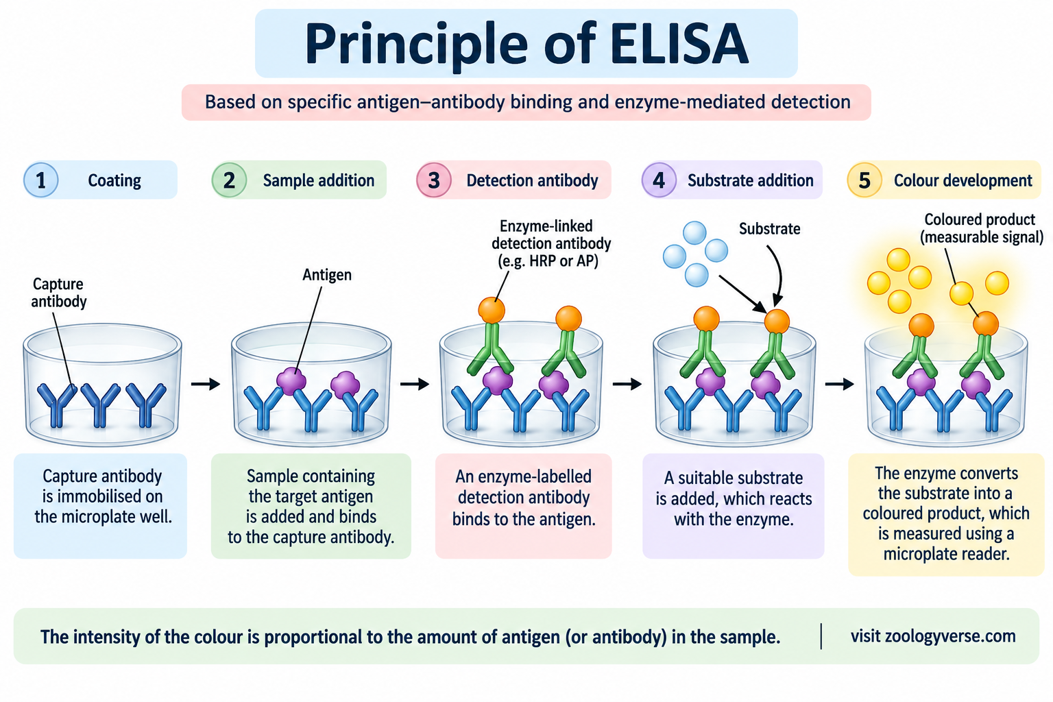
The principle of ELISA is based on the specific binding between an antigen and its corresponding antibody, together with enzyme-mediated detection. The assay uses an enzyme-linked reagent to generate a measurable signal, usually a colour change, after the addition of a suitable substrate.
In a typical ELISA, one component of the antigen–antibody reaction is attached to a solid surface, such as a microplate well. The sample is added, followed by appropriate detection reagents. After unbound substances are removed by washing, the enzyme–substrate reaction produces a signal that can be measured using a plate reader.
Steps Involved in the Principle of ELISA
1. Immobilisation of antigen or antibody
An antigen or capture antibody is attached to the surface of a microplate well. This provides a solid phase on which the immunological reaction takes place.
2. Addition of the sample
The sample is added to the well. If the target substance is present, it binds to the immobilised antigen or antibody, depending on the assay format.
3. Washing
The well is washed to remove unbound sample components. Effective washing helps reduce background signal and improves the reliability of the result.
4. Addition of enzyme-linked detection reagent
An enzyme-linked antibody or another suitable detection reagent binds to the target or to a previously bound antibody. The exact arrangement depends on whether the assay is direct, indirect, sandwich, or competitive.
5. Addition of the substrate
A suitable substrate is added. The enzyme catalyses a reaction that produces a detectable signal, commonly a coloured product.
6. Measurement of the signal
The colour intensity is measured using an ELISA plate reader, often by measuring absorbance at a specified wavelength. Quantitative assays generally use a standard curve to estimate the concentration of the target.
Role of Enzymes in ELISA
Commonly used enzymes include:
- Horseradish peroxidase (HRP): Frequently used with substrates such as TMB to produce a coloured reaction product.
- Alkaline phosphatase (AP): Used with suitable substrates to generate a measurable signal.
The choice of enzyme and substrate depends on the assay design and detection requirements.
Relationship Between Signal and Target Concentration
In many non-competitive ELISA formats, the measured signal increases as the amount of target increases, within the validated working range of the assay. In competitive ELISA, the signal commonly decreases as the target concentration increases.
Therefore, the result must be interpreted according to the assay format, controls, calibration standards, and manufacturer’s instructions.
Components and Requirements of ELISA
A successful ELISA depends on suitable reagents, laboratory equipment, and carefully controlled experimental conditions. Each component has a specific role in antigen–antibody binding, enzyme-based detection, or measurement of the final signal.
1. Microtitre Plate
A microtitre plate is the solid support used for the assay. It commonly contains 96 wells, although other formats are also available. The well surface allows antigens or capture antibodies to be immobilised so that the target can be detected.
2. Antigen or Capture Antibody
The immobilised component depends on the assay design.
- Antigen: May be attached to the well to detect antibodies in a sample.
- Capture antibody: Binds the target antigen in formats such as sandwich ELISA.
The binding component should have suitable specificity for the target being investigated.
3. Sample
The sample contains the antigen or antibody being investigated. Common sample types include serum, plasma, cell culture supernatants, and other biological fluids, depending on the validated assay.
4. Primary and Detection Antibodies
Antibodies recognise specific antigens and enable the target to be detected.
- A primary antibody may bind directly to the target antigen.
- A detection antibody recognises the target or another antibody already bound in the assay.
- In some formats, the detection antibody is directly linked to an enzyme. In others, an enzyme-linked secondary antibody is used.
Not every ELISA format requires separate primary and secondary antibodies.
5. Enzyme Conjugate
An enzyme conjugate contains an enzyme linked to an antibody or another detection reagent. Horseradish peroxidase (HRP) and alkaline phosphatase (AP) are commonly used enzymes.
The enzyme converts a suitable substrate into a detectable product.
6. Substrate and Chromogen
The substrate reacts with the enzyme to generate the detection signal. In colourimetric assays, this produces a coloured product.
For example, tetramethylbenzidine (TMB) is commonly used with HRP. The colour development and any stopping reagent must follow the specific assay protocol.
7. Washing Buffer
Washing buffer removes unbound materials between assay steps. Inadequate washing can produce high background signals, while overly harsh or inappropriate washing may interfere with specific binding.
8. Blocking Solution
A blocking solution covers unoccupied binding sites on the microplate surface. This helps prevent non-specific adsorption of proteins and reduces background signal.
Common blocking agents include bovine serum albumin (BSA) and casein, although the appropriate choice depends on the assay.
9. Standards and Controls
Standards and controls help establish whether the assay is performing correctly.
- Positive control: Helps confirm that the detection system can identify a known positive sample.
- Negative control: Helps assess background signal or non-specific reactions.
- Blank: Measures signal from relevant reagents without the target, according to the assay design.
- Calibration standards: Known concentrations used to construct a standard curve for quantitative assays.
10. Laboratory Equipment
The commonly required equipment includes:
- Micropipettes and suitable tips
- Microtitre plate and plate holder
- Plate washer or washing equipment
- ELISA plate reader
- Incubator, where required by the protocol
- Appropriate personal protective equipment
| Component | Main function |
|---|---|
| Microtitre plate | Provides a solid surface for the reaction |
| Antigen or capture antibody | Binds the target or provides assay specificity |
| Sample | Contains the target substance |
| Detection antibody | Enables specific detection |
| Enzyme conjugate | Converts substrate into a measurable signal |
| Substrate | Produces the detectable reaction product |
| Washing buffer | Removes unbound reagents |
| Blocking solution | Reduces non-specific binding |
| Standards and controls | Support calibration and result validation |
| Plate reader | Measures the assay signal |
Types of ELISA
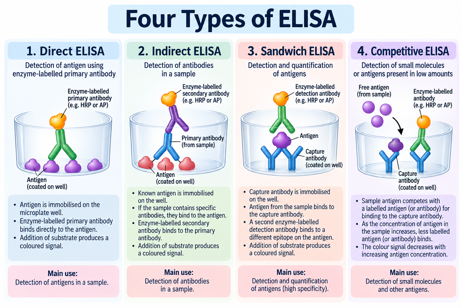
ELISA is classified into four major types according to how the antigen or antibody is immobilised and how the target substance is detected. These are direct ELISA, indirect ELISA, sandwich ELISA, and competitive ELISA.
Each format has specific advantages and is selected according to the target molecule, sample type, sensitivity requirements, and purpose of the test.
1. Direct ELISA
Direct ELISA detects an antigen using an enzyme-labelled primary antibody that binds directly to the target antigen.
Procedure:
- The antigen is immobilised on the microplate well.
- An enzyme-labelled primary antibody is added.
- The plate is washed to remove unbound antibody.
- The substrate is added to generate a measurable signal.
Advantages:
- Simple and relatively quick procedure.
- Fewer detection reagents are required.
- No secondary-antibody step is needed.
Limitations:
- Each primary antibody generally needs its own enzyme label.
- Signal amplification may be lower than in some indirect formats.
- Non-specific binding can affect results.
Applications: Detection of selected antigens in biological samples and laboratory research.
2. Indirect ELISA
Indirect ELISA is commonly used to detect specific antibodies in a sample. A known antigen is immobilised on the plate, and antibodies in the sample bind to it if they recognise that antigen. An enzyme-labelled secondary antibody then detects the bound sample antibodies.
Procedure:
- A known antigen is immobilised on the plate.
- The sample is added.
- Specific antibodies in the sample bind to the antigen.
- An enzyme-labelled secondary antibody is added.
- After washing, the substrate is added and the signal is measured.
Advantages:
- Can provide signal amplification because multiple secondary antibodies may bind to a primary antibody.
- One labelled secondary antibody may be used with several primary antibodies of the same species and antibody class.
- Useful for antibody detection.
Limitations:
- Non-specific secondary-antibody binding can increase background.
- Cross-reactivity may affect specificity.
- Additional incubation and washing steps may be required.
Applications: Detection of antibodies against particular pathogens or antigens, subject to the design and validation of the test.
3. Sandwich ELISA
Sandwich ELISA is used to detect antigens captured between two antibodies that recognise different epitopes on the same target. A capture antibody is attached to the plate, and a detection antibody binds to the captured antigen.
Procedure:
- A capture antibody is immobilised on the plate.
- The sample is added, allowing the target antigen to bind.
- The plate is washed to remove unbound material.
- A detection antibody is added.
- An enzyme-linked detection system is used, if the detection antibody is not already enzyme-labelled.
- The substrate is added and the signal is measured.
Advantages:
- Often provides high specificity because two recognition events are involved.
- Suitable for complex biological samples.
- Can be highly sensitive when the antibodies and assay conditions are well optimised.
Limitations:
- Requires compatible capture and detection antibodies.
- Assay development may be more complex.
- Some targets may be difficult to detect if suitable antibody-binding sites are unavailable.
Applications: Measurement of cytokines, hormones, and selected microbial or other protein antigens.
4. Competitive ELISA
Competitive ELISA measures a target by assessing how it competes with another antigen or labelled reagent for limited antibody-binding sites. In many common competitive formats, a greater amount of target in the sample results in a lower measured signal.
General procedure:
- The sample target is introduced into a competition system involving a specific antibody and a competing antigen or reagent.
- The target competes for antibody-binding sites.
- The resulting binding pattern determines how much labelled reagent remains associated with the solid phase.
- The enzyme–substrate reaction generates a measurable signal.
- The result is interpreted using a calibration curve.
The exact arrangement differs among competitive assay designs.
Advantages:
- Useful for measuring small molecules that may not have two suitable antibody-binding sites.
- Can be adapted to different target molecules.
- May be suitable for complex samples when appropriately validated.
Limitations:
- The signal commonly has an inverse relationship with target concentration.
- Interpretation and assay optimisation may be more complex.
- Reliable standards and controls are essential.
Applications: Measurement of selected hormones, drugs, toxins, and other small molecules.
Procedure of ELISA
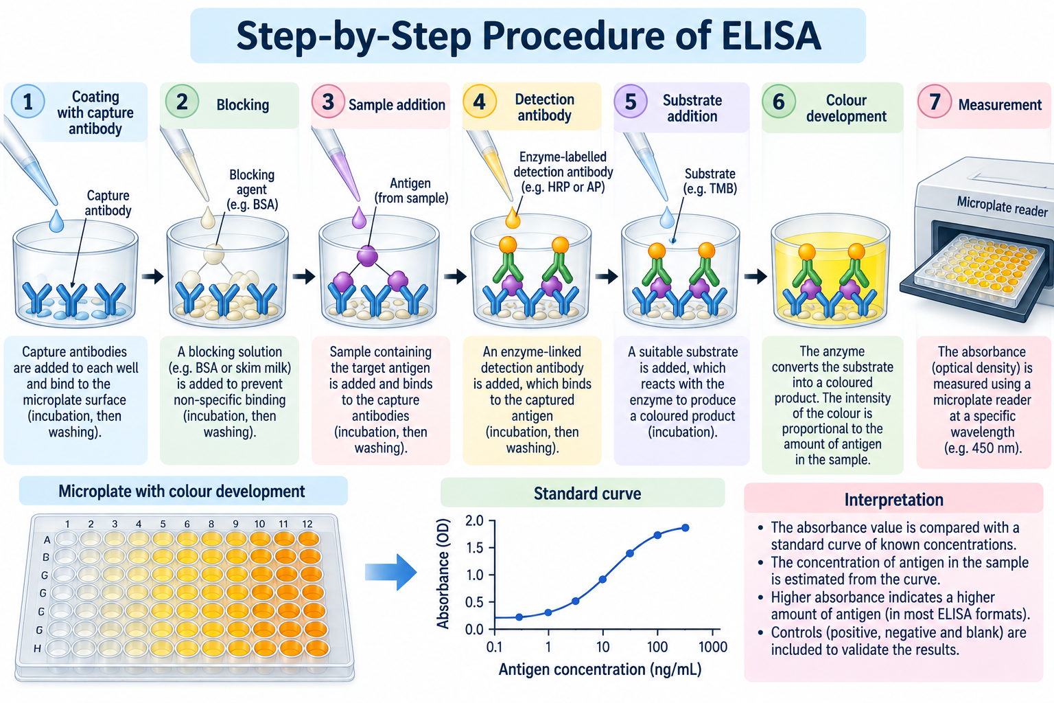
The procedure of ELISA involves a sequence of steps designed to capture the target substance, detect it using specific reagents, and generate a measurable signal. The exact procedure varies according to the assay format, sample type, and manufacturer’s instructions.
The following steps describe a general microplate-based procedure. A sandwich format is used as an example because it is commonly applied to antigen detection.
Step 1: Coating the Microplate
The wells of a suitable microtitre plate are coated with a specific capture antibody. The antibody attaches to the well surface and provides a binding site for the target antigen.
In other formats, an antigen or another binding reagent may be immobilised instead.
Step 2: Blocking
A blocking solution is added to cover unoccupied binding sites on the plate surface. This reduces non-specific adsorption of proteins and helps minimise background signal.
Step 3: Addition of the Sample
The biological sample is added to the wells. If the target antigen is present, it binds to the immobilised capture antibody.
Standards and appropriate controls are processed alongside the samples according to the assay protocol.
Step 4: Washing
The wells are washed to remove unbound sample components. Washing is essential because residual unbound material can interfere with detection and produce inaccurate results.
Step 5: Addition of the Detection Antibody
A detection antibody that recognises a different epitope on the target antigen is added. It binds to the captured antigen and forms an antibody–antigen–antibody complex.
Step 6: Addition of the Enzyme Conjugate
If the detection antibody is not directly enzyme-labelled, an enzyme-linked secondary detection reagent is added. The enzyme is commonly horseradish peroxidase (HRP) or alkaline phosphatase (AP).
Some assay designs combine the detection antibody and enzyme conjugate in a single reagent.
Step 7: Final Washing
The wells are washed again to remove unbound detection reagents. This step helps ensure that the subsequent signal mainly reflects specifically bound material.
Step 8: Addition of the Substrate
A suitable enzyme substrate is added. The enzyme catalyses a reaction that produces a measurable signal, often a colour change.
For example, HRP can react with tetramethylbenzidine (TMB) to produce a coloured product. The reaction conditions and any stopping reagent must follow the validated assay instructions.
Step 9: Measurement of the Signal
The reaction is measured using an ELISA plate reader at the appropriate wavelength. The measured absorbance is compared with the standards and controls.
In a quantitative assay, a calibration curve is used to estimate the concentration of the target substance. A qualitative assay may instead classify the result according to validated cut-off criteria.
Step 10: Interpretation and Reporting
The final result is interpreted according to the assay design, quality-control criteria, and manufacturer’s instructions. Results should not be interpreted from colour intensity alone without considering the controls and applicable cut-off values.
Flowchart of the General Procedure
Coating → Blocking → Sample addition → Washing → Detection antibody → Enzyme-linked reagent → Washing → Substrate addition → Signal measurement → Result interpretation
Note: The sequence and number of steps differ among direct, indirect, sandwich, and competitive formats. Follow the validated protocol for the specific assay being performed.
Interpretation of ELISA Results
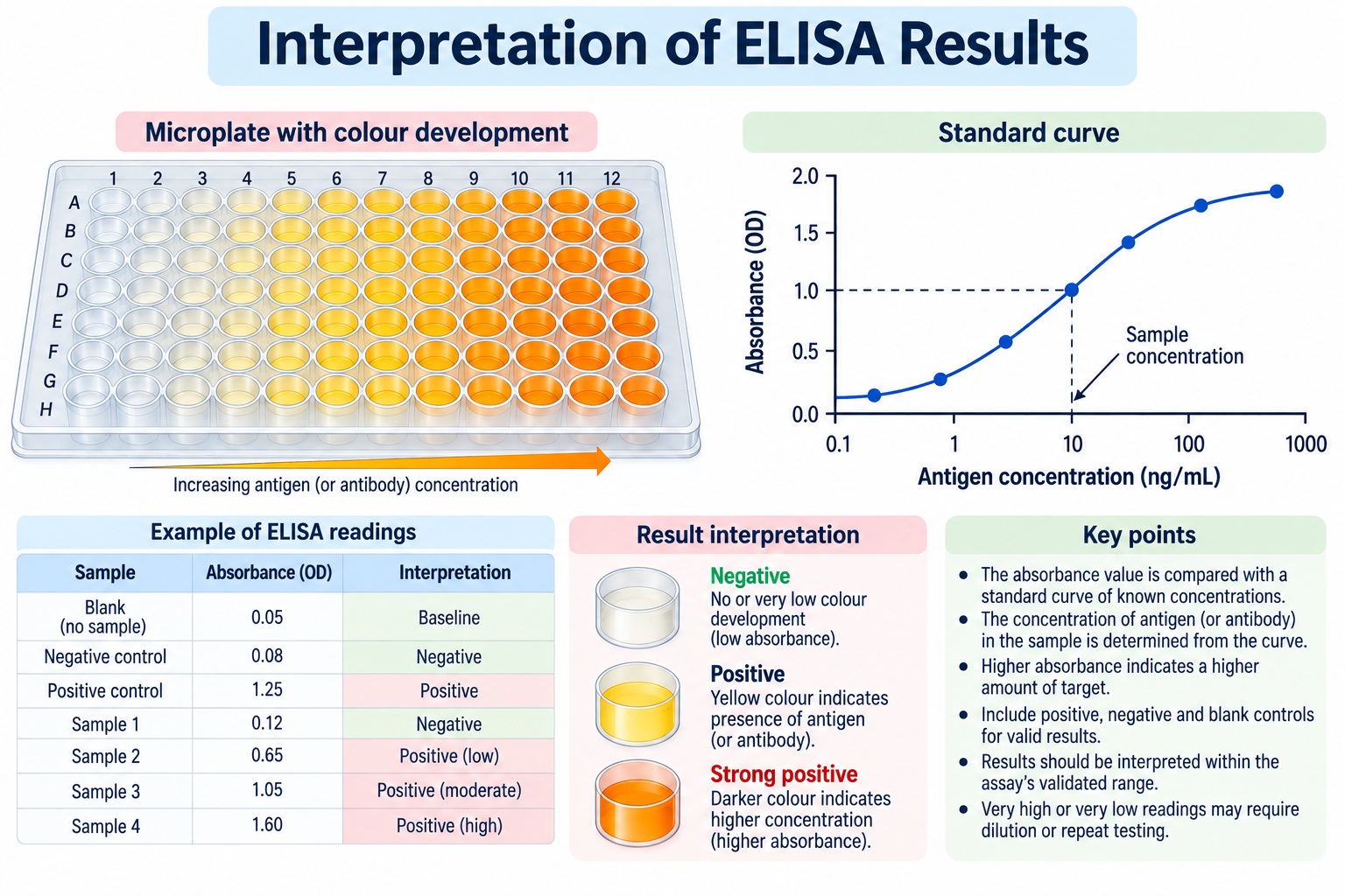
The interpretation of ELISA results involves evaluating the signal produced by the assay and comparing it with appropriate standards, controls, and validated decision criteria. Depending on the assay design, the result may indicate the presence or absence of a target substance or provide an estimate of its concentration.
1. Colour Development
In colourimetric assays, an enzyme reacts with its substrate to produce a coloured product. The intensity of the colour can provide information about the amount of target substance present.
In many non-competitive formats, a stronger signal indicates a higher target concentration within the validated measurement range. In competitive formats, the relationship is commonly inverse.
Colour intensity alone is not sufficient to determine a result; the assay’s controls and interpretation criteria must also be considered.
2. Measurement of Optical Density
The coloured product is measured using a microplate reader. The instrument records absorbance, often referred to as optical density (OD), at a wavelength specified by the assay protocol.
The measured absorbance provides a numerical signal that can be compared across wells and evaluated against standards or cut-off values.
3. Use of Standards and Controls
Standards and controls help determine whether the assay is functioning correctly.
- Positive control: Confirms that the assay can detect the expected target under the specified conditions.
- Negative control: Helps identify background signal or non-specific reactions.
- Blank: Measures signal associated with relevant reagents without the target, as defined by the protocol.
- Calibration standards: Known target concentrations used to construct a calibration curve for quantitative measurement.
If required controls fail their acceptance criteria, the results may be invalid and the assay may need to be repeated.
4. Qualitative Interpretation
A qualitative assay classifies a sample according to criteria established during assay validation.
Results may be reported as:
- Reactive or positive: The result meets the assay’s criteria for detection.
- Non-reactive or negative: The result does not meet the criteria for detection.
- Indeterminate or equivocal: The result falls within a range requiring additional evaluation, if such a category is defined by the assay.
A reactive result does not always establish a clinical diagnosis. Further testing may be necessary depending on the target and clinical context.
5. Quantitative Interpretation
Quantitative assays estimate the concentration of a target substance by comparing the sample signal with a calibration curve generated from standards of known concentrations.
The relationship between signal and concentration may be linear or non-linear. Many quantitative assays use a fitted curve, such as a four-parameter logistic model, over a defined measurement range.
Results outside the validated range may require dilution, repeat testing, or another procedure specified by the assay protocol.
6. Factors Affecting Results
Several factors can influence assay accuracy and reliability:
- Inadequate washing or non-specific binding
- Incorrect reagent preparation or storage
- Pipetting errors
- Improper incubation time or temperature
- Interfering substances in the sample
- Signal saturation or concentrations outside the measurement range
- Failure of positive or negative controls
Some assays may also be affected by high-dose hook effects, heterophile antibodies, or other matrix-related interference.
7. Clinical Significance
The meaning of a result depends on what the assay measures. For example, detecting antibodies against a pathogen is not necessarily equivalent to detecting the pathogen itself. Antibodies may reflect previous exposure, vaccination, or a response that requires interpretation alongside other clinical findings.
Therefore, results should be interpreted using the specific test’s validated criteria and, where relevant, the patient’s symptoms, history, and confirmatory tests.
Applications of ELISA
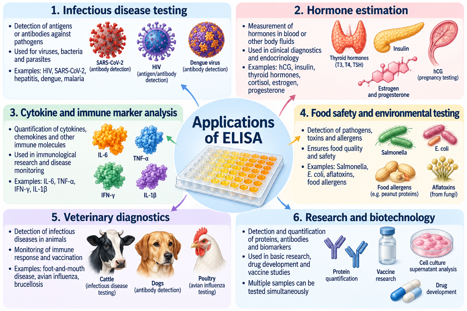
ELISA has a wide range of applications in clinical diagnosis, immunology, biomedical research, biotechnology, agriculture, and food safety. Its ability to detect specific antigens or antibodies and measure selected biological molecules makes it a valuable laboratory technique.
1. Diagnosis of Infectious Diseases
ELISA-based tests can detect antigens produced by pathogens or antibodies generated by the immune system in response to infection.
Examples include:
- HIV testing: Certain immunoassays detect HIV antigens, antibodies, or both as part of laboratory screening and diagnostic algorithms.
- Hepatitis B testing: ELISA-based methods can detect hepatitis B surface antigen (HBsAg) and selected antibodies associated with infection or immunity.
- Other infections: Appropriately designed immunoassays can help investigate infections such as dengue and selected parasitic diseases.
The interpretation of a result depends on the specific marker measured, the stage of infection, and the applicable testing guidelines.
2. Detection of Antibodies
Indirect ELISA is commonly used to identify antibodies that recognise a particular antigen. Such tests can help investigate previous exposure, immune responses, or selected immune-mediated conditions.
However, antibody detection does not always establish an active infection or guarantee protective immunity.
3. Hormone Estimation
ELISA-based immunoassays can measure hormones and other signalling molecules in biological samples.
Examples include:
- Human chorionic gonadotropin (hCG)
- Cortisol
- Insulin
- Selected reproductive and metabolic hormones
The assay format depends on the molecule being measured and the sensitivity required.
4. Detection of Allergens and IgE
Specific immunoassays can measure IgE directed against particular allergens. These tests may support the evaluation of allergic diseases when interpreted alongside clinical history and symptoms.
A positive allergen-specific IgE result indicates sensitisation; it does not by itself prove that exposure to that allergen causes clinical symptoms.
5. Measurement of Cytokines and Immune Proteins
ELISA is widely used in immunology research to measure cytokines, chemokines, and other proteins involved in immune responses.
Examples include interleukins, interferons, and tumour necrosis factor. These measurements can help researchers study inflammation, immune-cell activity, and responses to experimental treatments.
6. Cancer Research and Biomarker Detection
Selected immunoassays can measure tumour-associated proteins and other biomarkers in biological samples. Such measurements may contribute to research, disease monitoring, or particular clinical investigations.
However, not every cancer biomarker is suitable for screening, and an individual result cannot establish or exclude cancer without appropriate clinical evaluation.
7. Food Safety and Quality Control
ELISA-based methods can be used to detect specific food allergens, selected microbial antigens, and certain toxins or contaminants.
Examples include testing for traces of milk proteins, peanut proteins, or selected mycotoxins. The usefulness of a test depends on its validated detection limits, sample preparation requirements, and specificity.
8. Veterinary Medicine and Animal Research
ELISA is used in veterinary diagnostics and animal research to detect antibodies, antigens, hormones, and other biological markers. It can support investigations of infectious diseases, vaccination responses, reproductive physiology, and animal health.
9. Pharmaceutical and Biotechnology Research
In pharmaceutical research, immunoassays may be used to measure therapeutic proteins, antibodies, biomarkers, or immune responses. They can also help researchers evaluate biological samples during drug development and laboratory studies.
ELISA is a versatile analytical technique used to detect and quantify selected biological substances. Its results are most useful when the assay is properly validated and interpreted in the context of its intended application.
Advantages and Limitations of ELISA
ELISA is widely used because it combines specific antigen–antibody recognition with enzyme-based detection. However, its reliability depends on assay design, reagent quality, sample characteristics, and correct interpretation.
Advantages of ELISA
1. High specificity
The use of antibodies that recognise particular antigens allows the assay to detect selected target molecules. Specificity depends on antibody quality and the possibility of cross-reactivity.
2. High sensitivity
Appropriately designed assays can detect small amounts of an antigen or antibody. Sensitivity varies with the target, detection system, and assay conditions.
3. Quantitative capability
Many formats can estimate the concentration of a target using a calibration curve prepared from standards of known concentrations.
4. Relatively simple procedure
Microplate-based assays follow a structured sequence of coating or capture, incubation, washing, detection, and signal measurement.
5. High-throughput testing
Microtitre plates allow many samples and controls to be processed simultaneously, making the technique useful for routine laboratory testing and research.
6. No radioactive labels required
Unlike radioimmunoassays, conventional colourimetric ELISA uses enzyme-labelled reagents and suitable substrates rather than radioactive labels.
7. Versatility
Different assay formats can be adapted to detect antigens, antibodies, hormones, cytokines, and other biological molecules.
8. Automation compatibility
Many ELISA procedures can be performed using automated pipetting systems, plate washers, and plate readers, helping improve consistency and laboratory efficiency.
Limitations of ELISA
1. False-positive results
Cross-reactivity, non-specific binding, and interfering substances may produce a signal even when the target is absent.
2. False-negative results
A target may go undetected if its concentration is below the detection limit, if the relevant antibody does not recognise the target effectively, or if the sample interferes with the assay.
3. Dependence on antibody quality
The assay requires suitable recognition reagents. Poor antibody specificity, stability, or binding performance can reduce reliability.
4. Multiple procedural steps
Incubation, washing, reagent preparation, and timing must be controlled. Errors at any stage can affect the final measurement.
5. Limited measurement range
A quantitative assay is reliable only within its validated working range. Very high or very low concentrations may require additional procedures or another assay.
6. Potential interference
Heterophile antibodies, sample matrix effects, and certain interfering substances can affect some immunoassays. High concentrations of target may also cause a hook effect in certain sandwich formats.
7. Results may require confirmation
A positive or reactive result does not always establish a diagnosis. Some tests require confirmatory testing or interpretation alongside clinical findings.
Advantages and Limitations: Comparison Table
| Advantages | Limitations |
|---|---|
| Can provide high sensitivity | Sensitivity varies among assays |
| Specific antigen–antibody recognition | Cross-reactivity may occur |
| Can estimate target concentration | Requires suitable calibration for quantitative results |
| Processes many samples in parallel | Procedural errors can affect results |
| No radioactive labels in conventional formats | Requires appropriate enzymes and substrates |
| Adaptable to many biological targets | Depends on validated reagents and assay design |
| Can be automated | Equipment and quality-control requirements remain |
ELISA is a valuable analytical technique for immunological testing and biological research. Its major strengths are specificity, potential sensitivity, versatility, and suitability for processing multiple samples. Its limitations highlight the importance of validated procedures, appropriate controls, and careful interpretation of results.
ELISA vs Western Blot
ELISA and Western blot are immunological techniques used to detect specific biological molecules, particularly proteins and antibodies. Both rely on antigen–antibody recognition, but they differ in their basic principles, procedures, and applications.
ELISA commonly detects or quantifies a target through an enzyme-generated signal in a microplate. Western blot separates proteins by gel electrophoresis, transfers them to a membrane, and uses specific antibodies to detect proteins of interest.
Differences Between ELISA and Western Blot
| Feature | ELISA | Western Blot |
|---|---|---|
| Full form | Enzyme-Linked Immunosorbent Assay | Western blotting |
| Basic principle | Specific antigen–antibody binding with enzyme-based detection | Protein separation followed by antibody-based detection |
| Main purpose | Detection and, in suitable formats, quantification of antigens or antibodies | Detection of specific proteins, often with information about apparent molecular size |
| Solid support | Usually a microtitre plate | Usually a membrane such as nitrocellulose or PVDF |
| Separation of proteins | Generally not required | Proteins are separated by gel electrophoresis |
| Detection method | Commonly colourimetric; other detection systems are possible | Commonly chemiluminescent, colourimetric, or fluorescent |
| Sample processing | Often relatively straightforward | Usually involves gel preparation, electrophoresis, and transfer |
| Throughput | Well suited to processing many samples in parallel | Generally lower throughput for routine processing |
| Quantification | Commonly quantitative with suitable calibration | Can be semi-quantitative or quantitative when properly validated |
| Information about protein size | Usually not provided directly | Provides an estimate of apparent molecular size |
| Common applications | Antigen or antibody testing, hormone measurement, cytokine analysis | Protein identification, expression analysis, and investigation of protein size or processing |
Advantages of ELISA Compared with Western Blot
- It is generally easier to adapt for high-throughput sample testing.
- It can estimate target concentrations using a calibration curve.
- Many microplate formats are compatible with automated equipment.
- It often requires less sample-processing time than a complete Western blot workflow.
Advantages of Western Blot Compared with ELISA
- It separates proteins by electrophoretic mobility before detection.
- It provides information about the apparent molecular size of a detected protein.
- The banding pattern can help researchers investigate protein expression, degradation, or unexpected protein species.
However, a Western blot band does not by itself prove the identity of a protein, and both techniques can be affected by antibody specificity and other experimental limitations.
Which Technique Is Better?
Neither technique is universally better. ELISA is often preferred when many samples must be screened or a target concentration needs to be measured efficiently. Western blot is useful when protein separation and information about apparent molecular size are important.
The choice depends on the research question, the sample, the target molecule, and the required evidence. In some investigations, both methods may be used because they provide complementary information.
Frequently Asked Questions (FAQs) About ELISA
What is ELISA?
What is the basic principle of ELISA?
What are the four main types of ELISA?
Which enzyme is commonly used in ELISA?
What is the role of the substrate?
What is the difference between direct and indirect ELISA?
Why is sandwich ELISA often highly specific?
What is competitive ELISA used for?
What is the role of washing in ELISA?
Can ELISA provide quantitative results?
Is a positive ELISA result always proof of disease?
What are the main applications of ELISA?
Key Takeaways
- ELISA stands for Enzyme-Linked Immunosorbent Assay.
- It is an immunological technique used to detect or quantify specific antigens or antibodies.
- The principle is based on specific antigen–antibody binding and enzyme-mediated signal generation.
- The assay commonly uses microtitre plates as a solid support.
- Enzymes such as horseradish peroxidase (HRP) and alkaline phosphatase (AP) are commonly used.
- Suitable substrates produce a measurable signal, often a colour change.
- A plate reader measures absorbance in colourimetric assays.
- The four major types are direct, indirect, sandwich, and competitive ELISA.
- Direct ELISA uses an enzyme-labelled primary antibody to detect an antigen.
- Indirect ELISA commonly detects antibodies using an enzyme-labelled secondary antibody.
- Sandwich ELISA captures the target antigen between capture and detection antibodies.
- Competitive ELISA involves competition for antibody-binding sites; its signal commonly decreases as target concentration increases.
- Blocking reduces non-specific binding to the plate surface.
- Washing removes unbound reagents and helps reduce background signal.
- Positive controls, negative controls, blanks, and standards help assess assay performance.
- Quantitative assays use a calibration curve to estimate target concentration.
- Applications include infectious disease testing, hormone estimation, cytokine measurement, food safety, and biomedical research.
- Advantages include potential high sensitivity, specificity, versatility, and high-throughput testing.
- Limitations include possible false-positive and false-negative results, reagent dependence, and assay interference.
- Western blot differs from ELISA because it separates proteins by gel electrophoresis before antibody-based detection.
Conclusion
ELISA (Enzyme-Linked Immunosorbent Assay) is an important immunological technique used to detect and quantify specific antigens or antibodies. It combines the specificity of antigen–antibody interactions with enzyme-mediated signal generation, commonly producing a measurable colour change.
The four major formats—direct, indirect, sandwich, and competitive ELISA—differ in their detection mechanisms and applications. The technique is widely used in infectious disease testing, hormone estimation, immunological research, food safety, and biotechnology.
Its sensitivity, versatility, and suitability for processing multiple samples make it valuable in modern laboratories. However, reliable results depend on appropriate assay design, validated procedures, suitable controls, and correct interpretation. Understanding the principle, procedure, types, advantages, and limitations of ELISA is therefore essential for students of zoology, immunology, biotechnology, and biomedical sciences.
References
- Engvall, E., and Perlmann, P. (1971). Enzyme-linked immunosorbent assay (ELISA). Immunochemistry, 8(9), 871–874.
https://doi.org/10.1016/0019-2791(71)90454-X - Lequin, R. M. (2005). Enzyme immunoassay (EIA)/enzyme-linked immunosorbent assay (ELISA). Clinical Chemistry, 51(12), 2415–2418.
https://doi.org/10.1373/clinchem.2005.051532 - Crowther, J. R. (2009). The ELISA Guidebook (2nd ed.). Humana Press.
https://doi.org/10.1007/978-1-60327-254-4 - Aydin, S. (2015). A short history, principles, and types of ELISA, and our laboratory experience with peptide/protein analyses using ELISA. Peptides, 72, 4–15.
https://doi.org/10.1016/j.peptides.2015.04.012 - Wild, D. (ed.). (2013). The Immunoassay Handbook: Theory and Applications of Ligand Binding, ELISA and Related Techniques (4th ed.). Elsevier.
https://www.sciencedirect.com/book/9780080970370/the-immunoassay-handbook - Thermo Fisher Scientific. ELISA Learning Center. Technical resources on ELISA principles, reagents, and assay formats.
https://www.thermofisher.com/ - Abcam. ELISA Protocol and Technical Resources. Guidance on assay preparation, washing, detection, and result interpretation.
https://www.abcam.com/ - National Center for Biotechnology Information (NCBI), PubMed. Research literature on enzyme-linked immunosorbent assays.
https://pubmed.ncbi.nlm.nih.gov/?term=enzyme-linked+immunosorbent+assay
Discover more from Zoologyverse
Subscribe to get the latest posts sent to your email.