
Lepus is a genus of mammals belonging to the family Leporidae and order Lagomorpha. The male reproductive system of lepus is specialized for the production, maturation, storage, and transport of sperm and for the delivery of semen into the female reproductive tract.
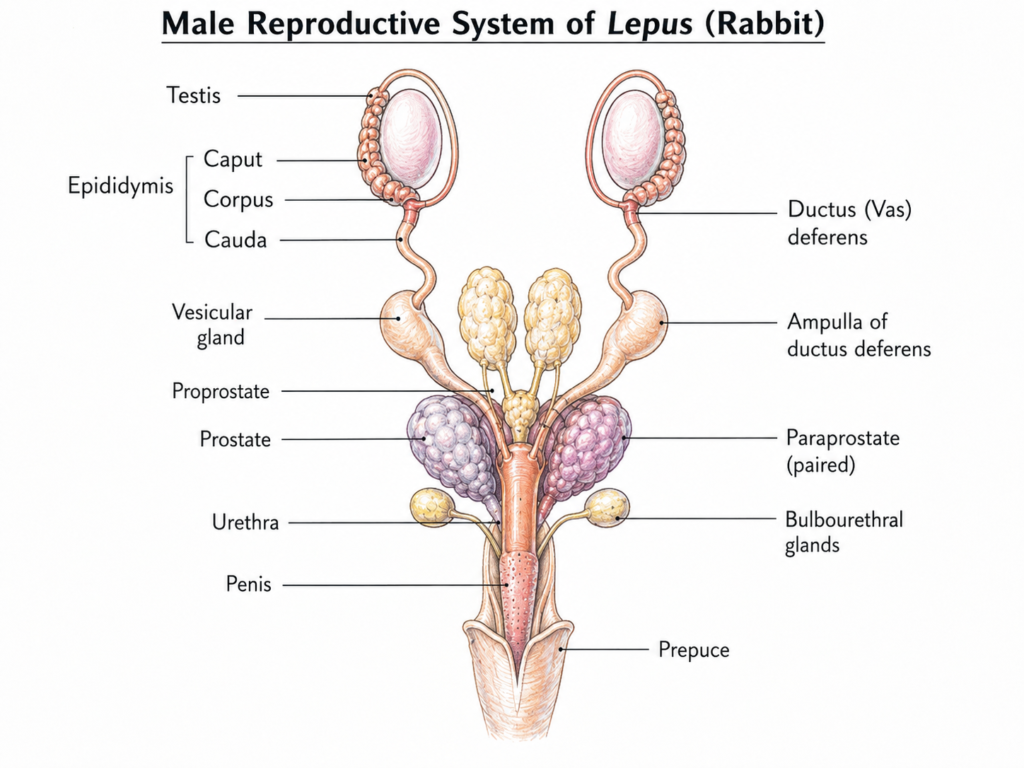
The male reproductive system of Lepus consists of a pair of testes, epididymides, vasa deferentia, accessory reproductive glands, urethra, penis, and prepuce. The testes are responsible for sperm production and androgen secretion, while the ducts and accessory glands modify and transport the sperm-containing secretion.
Taxonomic note: Detailed anatomical descriptions of the male reproductive system are particularly well documented in the domestic rabbit (Oryctolagus cuniculus), a close relative of Lepus. Because Lepus and Oryctolagus are both leporids, their general male reproductive organization is highly comparable; however, exact glandular morphology and measurements should not be assumed to be identical between the two genera.
General Organization of the Male Reproductive System of Lepus
The male reproductive system of Lepus follows the general mammalian pattern and is organized into primary reproductive organs, sperm-conducting ducts, accessory reproductive glands, and copulatory organs.
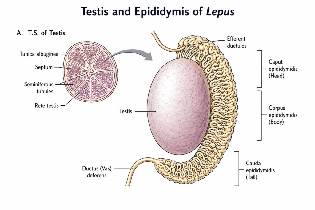
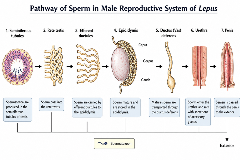
The paired testes are the primary reproductive organs. They produce spermatozoa and the male sex hormone testosterone. Sperm produced within the seminiferous tubules passes through the rete testis and efferent ductules before reaching the epididymis, where it undergoes maturation and is stored.
The mature sperm then travels through the ductus deferens toward the pelvic reproductive tract. Near its terminal portion, the ductus deferens forms an ampulla. Secretions from the accessory reproductive glands are added to the sperm before the resulting semen passes through the urethra and penis.
Detailed anatomical studies of the male domestic rabbit, Oryctolagus cuniculus, describe a particularly specialized accessory-gland system consisting of the vesicular gland, proprostate, prostate, paraprostate, and bulbourethral glands. Because Oryctolagus is a close relative of Lepus, this anatomy provides useful comparative information, but rabbit-specific observations should not automatically be presented as measurements or species-specific findings for every Lepus species.
Main Components of the Male Reproductive System of Lepus
The male reproductive system can be divided into the following major parts:
- Testes
- Epididymides
- Vasa deferentia
- Accessory reproductive glands
- Urethra
- Penis and prepuce
Testes
The testes are the paired primary male reproductive organs. Their two major functions are spermatogenesis and secretion of androgens, principally testosterone.
Each testis is enclosed by connective tissue and contains numerous highly coiled seminiferous tubules. These tubules are the site of sperm production. The developing germ cells are supported by Sertoli cells, while Leydig cells, located in the interstitial tissue between the seminiferous tubules, produce testosterone.
The sperm formed in the seminiferous tubules does not immediately enter the epididymis. It passes through a series of conducting channels:
Seminiferous tubules → tubuli recti → rete testis → efferent ductules → epididymis
The rete testis forms a network of channels that collects sperm from the seminiferous tubules. The efferent ductules then carry the sperm toward the head of the epididymis.
Thus, the testis performs both an exocrine function, by producing spermatozoa, and an endocrine function, by secreting testosterone.
Structure of the Testis
Each testis is enclosed by a dense connective-tissue capsule called the tunica albuginea. Internally, connective-tissue septa divide the testis into numerous lobules.
Each lobule contains highly coiled seminiferous tubules, where spermatogenesis takes place.
The general pathway of sperm formation and transport is:
Seminiferous tubules → rete testis → efferent ductules → epididymis
The seminiferous tubules contain germ cells at different stages of development along with Sertoli cells. Between the seminiferous tubules are groups of Leydig cells, which secrete testosterone.
Epididymis
A highly coiled epididymis is attached to the testis. It is divided into three major regions:
- Caput epididymidis – head
- Corpus epididymidis – body
- Cauda epididymidis – tail
The epididymis receives sperm from the testis through the efferent ductules.
Its major functions are:
- maturation of spermatozoa,
- concentration of sperm,
- temporary storage of sperm, particularly in the caudal region, and
- transport of sperm toward the ductus deferens.
The epididymis is a long, highly coiled duct closely associated with each testis. It is divided into three principal regions:
- Caput epididymidis — head
- Corpus epididymidis — body
- Cauda epididymidis — tail
The efferent ductules connect the rete testis with the caput epididymidis. As sperm passes through the epididymal duct, it undergoes important post-testicular maturation and becomes progressively capable of normal motility and fertilizing activity.
The cauda epididymidis serves as an important region for storage of mature sperm before ejaculation. From the caudal region, the sperm enters the ductus deferens.
The epididymis therefore acts not simply as a connecting duct but as an important organ for sperm maturation, concentration, storage, and transport.
Ductus Deferens
The ductus deferens, also called the vas deferens, is a paired muscular duct that transports sperm from the cauda epididymidis toward the pelvic reproductive tract.
Its muscular wall enables peristaltic contractions that propel sperm during ejaculation. Near its terminal portion, the ductus deferens becomes enlarged to form the ampulla of the ductus deferens.
In detailed rabbit anatomy, the ampullae lie near the vesicular gland and communicate with the ejaculatory ducts. The terminal ducts ultimately open into the pelvic urethral region.
The pathway can therefore be summarized as:
Cauda epididymidis → ductus deferens → ampulla → terminal reproductive ducts → urethra
Accessory Reproductive Glands
The accessory reproductive glands contribute secretions to the sperm and form the fluid portion of semen. In leporids, this group is relatively complex.
The accessory glands described in rabbit anatomy include:
- Vesicular glands
- Proprostate
- Prostate
- Paraprostate
- Bulbourethral glands
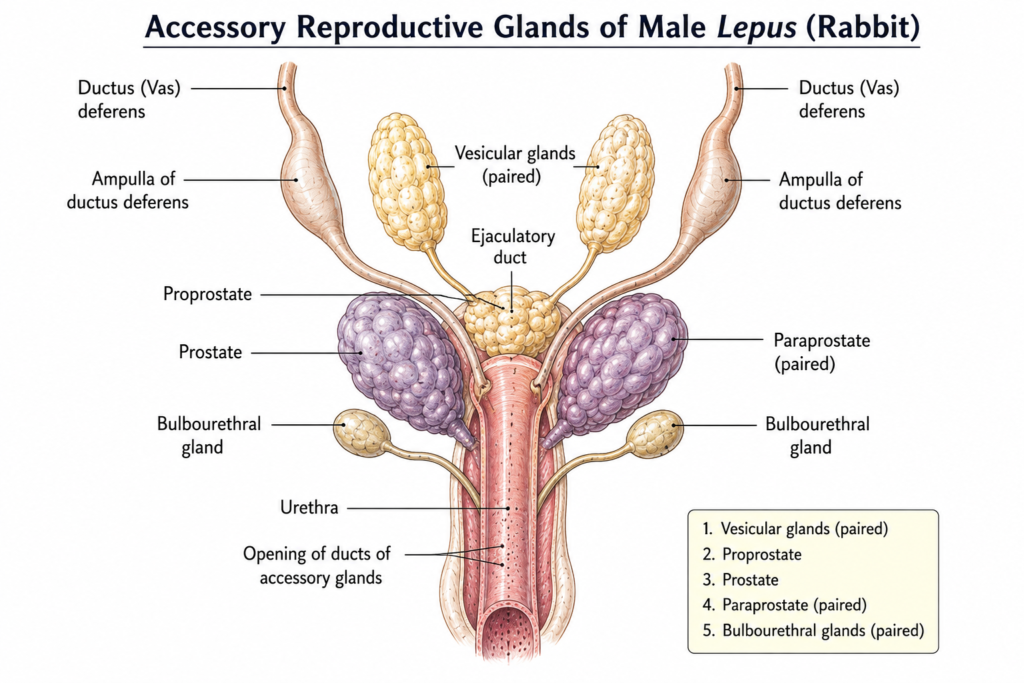
The terminology of these glands has historically varied, which is why older zoology texts may use different names for some of them. Detailed anatomical work specifically distinguished the vesicular, proprostate, prostate, paraprostate, and bulbourethral glands.
A. Vesicular Glands
The vesicular glands are paired accessory glands associated with the region of the urinary bladder and pelvic urethra.
They produce a secretion that contributes substantially to seminal fluid. In rabbits, the vesicular gland is a sac-like structure whose secretion can accumulate within its lumen. Its duct communicates with the urethral region through the ejaculatory duct.
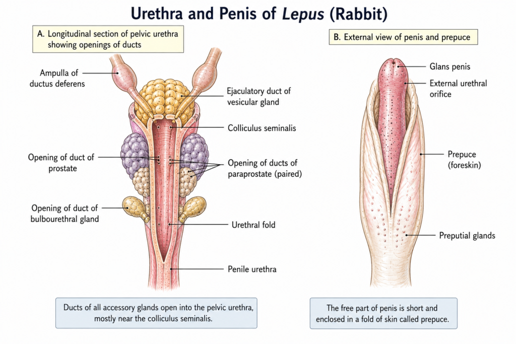
The vesicular gland is a specialized accessory gland associated with the dorsal aspect of the pelvic reproductive tract. In the domestic rabbit, it is a sac-like gland with a lumen that contains its secretion. Its terminal portion forms an ejaculatory duct, which communicates with the urethra near the colliculus seminalis.
B. Proprostate
The proprostate is a specialized component of the rabbit/leporid prostate complex. It lies in the pelvic region near the other accessory glands and contributes glandular secretion to the reproductive tract.
Because of historical differences in terminology, the proprostate has sometimes been incorrectly identified or grouped with other accessory glands in older descriptions. Modern anatomical studies recognize it as a distinct component of the rabbit prostate complex.
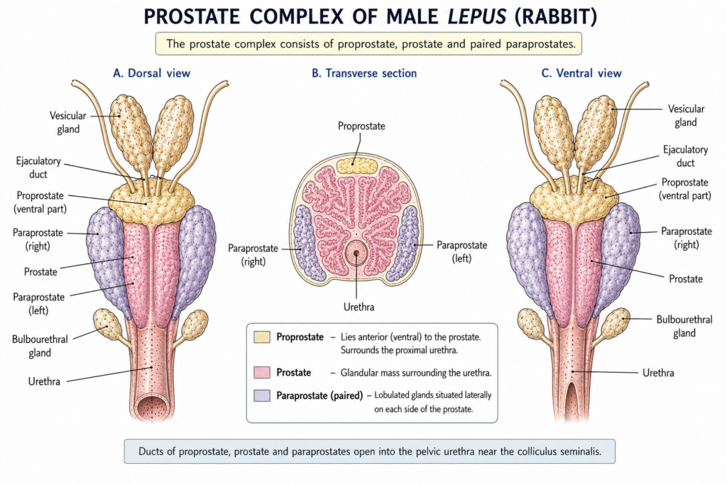
The proprostate is a distinct accessory gland situated cranial to the prostate in the rabbit. It possesses its own excretory ducts and contributes glandular secretion to the reproductive tract.
C. Prostate Gland
The prostate gland is situated around the pelvic part of the urethra and contributes glandular fluid to semen.
In rabbits, the prostate complex is more elaborate than the simple prostate often shown in generalized mammalian diagrams. Studies describe the prostate complex as including the proprostate, prostate proper, and paraprostate.
The prostate is a glandular organ associated with the pelvic urethra. In the rabbit, it is anatomically distinct from the proprostate and has its own excretory ducts.
D. Paraprostate
The paraprostate consists of accessory glandular tissue associated with the prostate complex. It is located laterally in relation to the other components of the complex and has its own ducts opening into the urethral region.
The number and arrangement of paraprostatic lobes can vary among individuals, which has been documented in detailed histological studies of rabbits.
The paraprostate consists of small accessory glandular structures associated with the dorsolateral region of the urethra. Their number and lobular arrangement can show considerable individual variation in rabbits.
E. Bulbourethral Glands
The paired bulbourethral glands are located caudally in relation to the prostate complex and are associated with the pelvic/perineal part of the urethra.
They produce a glandular secretion that contributes to the seminal fluid and helps prepare the urethral passage during reproductive activity.
In rabbit anatomy, the bulbourethral glands are paired and located dorsal to the urethra and caudal to the prostate/proprostate region. Their ducts open into the urethra.
The paired bulbourethral glands are situated caudal to the prostate/proprostate region and dorsal to the urethra. Their ducts open into the urethra. In rabbit anatomy, these glands are bilobed and are associated with the bulbourethral muscle.
Urethra
The urethra serves as a common passage for the urinary and reproductive systems in the male.
During reproduction, sperm and accessory gland secretions enter the pelvic urethra and are carried toward the penis.
A specialized structure called the colliculus seminalis occurs in the pelvic urethra. In the rabbit, the ejaculatory duct associated with the vesicular gland opens near this structure.
Thus, the urethra provides the final common pathway for the passage of semen to the exterior.
Penis
The penis is the copulatory organ of the male. It provides the passage through which semen is discharged into the female reproductive tract.
The penis contains erectile tissues and is connected posteriorly with the pelvic urethra. In the rabbit, the free portion of the penis is relatively short and the glans is small.
The penis is normally enclosed within a fold of skin called the prepuce.
Prepuce
The prepuce, or foreskin, is a skin fold surrounding and protecting the free portion of the penis.
In rabbits, small preputial glands are also associated with the preputial region.
Position and Arrangement of the Reproductive Organs
The male reproductive organs are distributed between the inguinal/scrotal region and the pelvic cavity.
The testes are associated with the scrotal region, while the epididymides remain closely attached to them. The ductus deferentia extend from the epididymides toward the pelvic cavity.
Within the pelvic region, the terminal portions of the ductus deferentia and the accessory reproductive glands are closely associated with the urinary bladder, urethra, and surrounding pelvic tissues.
In detailed rabbit anatomical studies, the vesicular gland, proprostate, prostate, paraprostate, and bulbourethral glands are positioned around the pelvic part of the urethra. This close anatomical relationship is important because their ducts empty into the urethral reproductive pathway.
Pathway of Sperm
Complete Pathway of Sperm
The movement of sperm through the male reproductive system can be represented as:
Seminiferous tubules
↓
Tubuli recti
↓
Rete testis
↓
Efferent ductules
↓
Caput epididymidis
↓
Corpus epididymidis
↓
Cauda epididymidis
↓
Ductus deferens
↓
Ampulla of ductus deferens
↓
Urethra
↓
Penis
↓
Exterior
During passage through the reproductive tract, sperm undergoes maturation in the epididymis and mixes with secretions from the accessory reproductive glands. The resulting semen is transported through the urethra and discharged through the penis.
Functional Organization
The male reproductive system of Lepus can therefore be understood according to the function of each component:
| Structure | Principal function |
|---|---|
| Testes | Sperm production and testosterone secretion |
| Seminiferous tubules | Site of spermatogenesis |
| Epididymis | Sperm maturation, concentration and storage |
| Ductus deferens | Transport of sperm |
| Ampulla of ductus deferens | Enlarged terminal portion associated with sperm transport and glandular tissue |
| Vesicular glands | Production of seminal secretion |
| Proprostate | Accessory glandular secretion |
| Prostate | Production of prostatic secretion |
| Paraprostate | Accessory glandular secretion |
Key Takeaways
- The male reproductive system of Lepus consists of paired testes, epididymides, ductus deferentia, accessory reproductive glands, urethra, and penis.
- The testes are the primary reproductive organs and produce spermatozoa and testosterone.
- The epididymis is divided into caput, corpus, and cauda and is the main site of sperm maturation and storage.
- The ductus deferens transports sperm from the epididymis toward the pelvic reproductive tract.
- The accessory gland system is complex and includes vesicular glands, proprostate, prostate, paraprostate, and bulbourethral glands in the closely related rabbit.
- The urethra serves as the common passage for semen and urine.
- The penis acts as the copulatory organ and delivers semen to the female reproductive tract.
- Because detailed anatomical literature is more extensive for Oryctolagus cuniculus, species-specific details should be distinguished from generalized leporid anatomy.
Frequently Asked Questions
What are the primary reproductive organs in male Lepus?
The paired testes are the primary male reproductive organs. They produce spermatozoa and secrete testosterone.
What is the function of the epididymis?
The epididymis is responsible mainly for sperm maturation, concentration, storage, and transport.
Which accessory reproductive glands occur in the rabbit/leporid reproductive system?
Detailed rabbit studies recognize the vesicular glands, proprostate, prostate, paraprostate, and bulbourethral glands.
What is the function of the ductus deferens?
The ductus deferens is a muscular tube that transports mature sperm from the cauda epididymidis toward the urethral reproductive tract.
Does the male rabbit have a prostate?
Yes. The prostate is part of a relatively complex accessory gland system in rabbits and is associated with the proprostate and paraprostate.
What is the role of the penis?
The penis is the male copulatory organ and provides the terminal passage through which semen is discharged.
Conclusion
The male reproductive system of Lepus is a highly organized system adapted for efficient sperm production, maturation, transport, and deposition. Its principal components are the testes, epididymides, ductus deferentia, accessory reproductive glands, urethra, and penis. The accessory gland complex is particularly noteworthy because leporid reproductive anatomy includes specialized structures such as the proprostate and paraprostate in addition to the vesicular, prostate, and bulbourethral glands. Detailed anatomical studies of rabbits provide an important comparative basis for understanding this system, while species-specific differences should be considered when applying rabbit anatomy directly to Lepus.
Scientific Note on Lepus and Rabbit Anatomy
Detailed anatomical literature on the male reproductive tract is considerably more extensive for the domestic rabbit, Oryctolagus cuniculus, than for species of Lepus. The two genera belong to the family Leporidae and share the general organization of the male reproductive system. Therefore, rabbit anatomical studies are useful for explaining specialized leporid structures, particularly the accessory reproductive glands. However, measurements, histological details, gland dimensions, and other species-specific observations obtained from Oryctolagus cuniculus should not be interpreted automatically as exact measurements or characteristics of Lepus. Where appropriate, this article uses rabbit anatomy as comparative evidence rather than claiming that every detailed observation has been directly established in Lepus.
References
- Holtz, W. & Foote, R. H. (1978). The anatomy of the reproductive system in male Dutch rabbits (Oryctolagus cuniculus) with special emphasis on the accessory sex glands. Journal of Morphology, 158, 1–20.
- Urinary Catheterization of Male Rabbits: A New Technique and a Review of Urogenital Anatomy. PMC.
- Accessory Genital Glands in the New Zealand White Rabbit: A Morphometrical and Histological Study. PMC.
- Bozbiyik et al. (2023). Investigation of male genital system anatomy in the New Zealand rabbit (Oryctolagus cuniculus L.). Anatomia, Histologia, Embryologia.
Discover more from Zoologyverse
Subscribe to get the latest posts sent to your email.