Amphioxus slides are essential teaching tools used in zoology and comparative anatomy to study the structural features of Branchiostoma (Amphioxus), a primitive chordate. These prepared slides display key characteristics such as the notochord, dorsal nerve cord, myotomes, pharyngeal gill slits, and segmented musculature. Students and researchers use Amphioxus slides to observe the organism’s simple yet significant body plan, which offers crucial insight into the evolution of vertebrates. Because Amphioxus occupies an important position between invertebrates and vertebrates, these slides help learners understand early chordate organization and fundamental concepts of developmental and evolutionary biology.
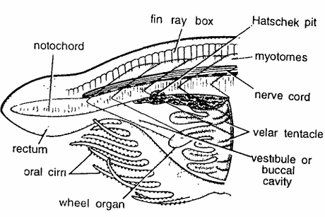
Amphioxus Slides : V.L.S. Anterior Region
Comments
- The vertical longitudinal section shows buccal cirri, wheel organ, velum and some pharyngeal region.
- In a carmine stained section, prominent dorsal structures are fin rays, notochord and nerve cord.
- Dorsal fin is low, continuous and supported by fin rays.
- Nerve cord or spinal cord lies just above the notochord.
- It contains anterior and posterior pigmented spots and anteriorly swollen as cerebral vesicle.
- Notochord lies just above the nerve cord forming axial skeletal rod.
- It extends antero posteriorly. Anterior end projects as the rostrum.
- Important ventral structures are oral hood, vestibule, wheel organ, pharynx and atrium.
- Oral hood is clearly seen with oral cirri, which help during feeding by turning inwards to prevent sand particles from passing into buccal cavity.
- Oral hood guards the vestibule or buccal cavity.
- At the hinder wall of vestibule lies a vertical partition called velum with velar tentacles.
- In front of velum is a peculiar wheel organ which helps in driving a current of water loaded with food particles into the mouth.
Identification: Since the above section has oral cirri and all above features, hence it is Amphioxus V.L.S. region.
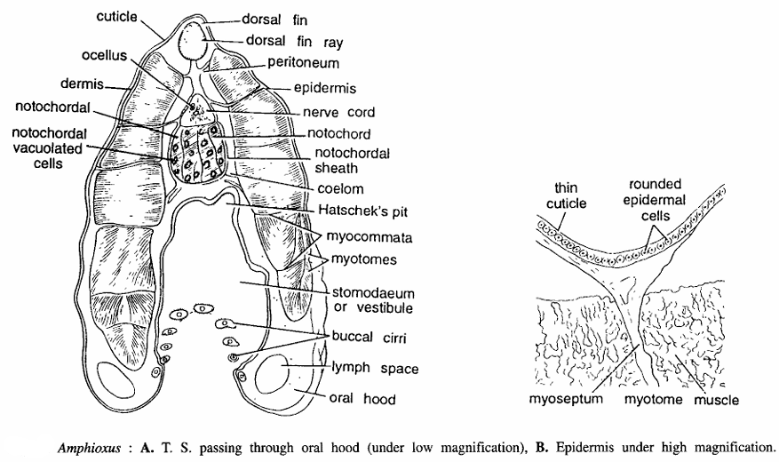
Amphioxus Slides : T.S. Passing Through Oral Hood
Comments
A. Under low magnification: (10 X eye-piece; 4 X objective).
- At the anterior end of Amphioxus is a mid-ventral opening encircled by frilled membrane, called oral hood.
- The T.S. Passing through the oral hood shows body wall, dorsal fin ray, nerve cord, notochord, vestibule and oral hood, etc.
- Body wall is composed of epidermis, dermis or cutis and muscle layer.
- Epidermis is covered by a non-pigmented and iridescent cuticle. Unlike other chordates, the Amphioxus epidermis is very thin.
- Dermis is indistinct. Below epidermis and dermis is a thick longitudinal muscle layer.
- The cut segmental blocks or myotomes are very distinct, separated by myosepta.
- The muscle fibers in anterior half section are directed upwards while in posterior half, backwards. Below muscle layer is coelom. Dorsally below the epidermis is a dorsal fin ray.
- Dorsal tubulated glandular nerve cord having a central canal or neurocoel and below it notochord are clearly seen.
- The notochord is composed of chordal or fibrous sheath, which encloses vacuolated notochordal cells filled with homogeneous liquid.
- Ventrally, section shows a large stomodaeum, oral hood and cut part of buccal cirri in a circular manner.
- Oral hood contains lymph spaces. Dorsal wall of buccal cavity has a sensory Hatscheck’s groove.
Under high magnification: (10 X eye-piece; 40 X objective).
- Epidermis is vary clearly seen under this magnification.
- It is composed of single layered rounded epithelial cells with some chemoreceptor cells and unicellular gland cells covered by thin cuticle. The myosepta are continuous with epidermis and visceral layer.
- Myotome muscles and myoseptum are seen.
Identification: Since the section has buccal cirri and all above features, hence it is T.S. passing through oral hood of Amphioxus.
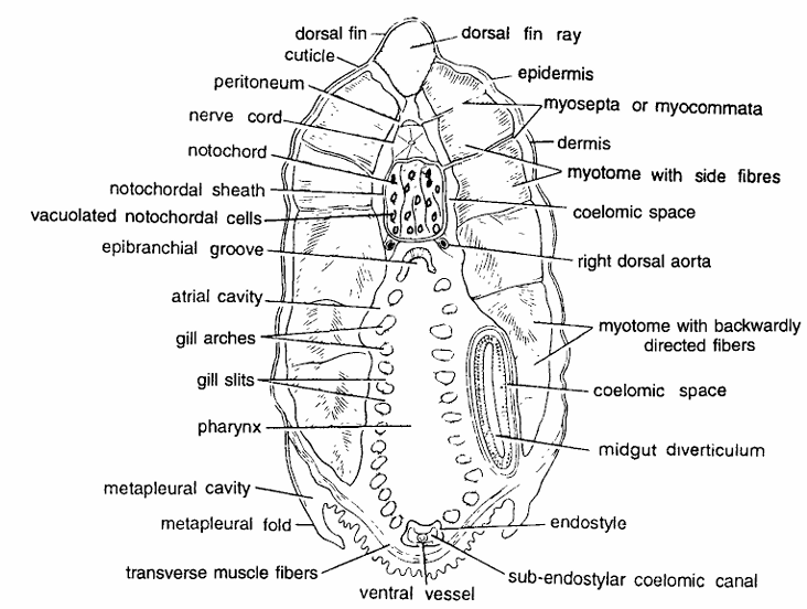
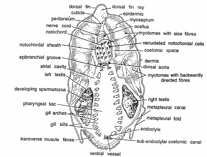
Amphioxus Slides : T.S. Passing Through Pharynx
Comments
- Pharynx is a large elongated, sac-like respiratory and digestive organ, extending from behind velum upto the intestine.
- T.S. passing through anterior pharynx shows body wall layers, dorsal rm ray, nerve cord, notochord, large cut pharynx with endostyle and metapleural folds.
- Body wall is composed of cuticle, epidermis, dermis and muscle layer.
- Cuticle and epidermis are thin-layered and indistinguishable. Below epidermis the dermis is also thin-layered.
- More than three-fourth of the section from dorsal side contains thick; cut, segmental muscle bundles or myotomes separated by transverse myosepta.
- The first three myotomes have side muscle fibres whlle in posterior, half the muscle fibers are backwardly directed.
- Dorsally, just beneath epidermis, is the dorsal fin ray. Below dorsal fin ray is nerve cord and beneath nerve cord is notochord.
- Notochord is surrounded by notochordal sheath and filled with vacuolated notochordal cells.
- Ventral half of the section contains the large pharynx surrounded by atrial cavity and perforated by gill slits.
- It contains longitudinal rows of cilia in the form of an epipharyngeal groove mid-dorsally and an endostyle enclosing an endostylar canal, midventrally.
- The ciliated grooves direct food material towards oesophagus.
- The sides of the pharyngeal cavity contain several gill arches.
- Pharynx is adapted for ciliary feeding.
- Two metapleural folds with metapleural cavity are seen posteriorly.
- In some sections through pharynx, midgut diverticulum or liver is also seen.
- Other structures seen are dorsal aorta, coelomic spaces, gill arches, ventral vessel and transverse muscle fibers.
Identification: Since this section shows epipharyngeal groove, gill slits and all above features, hence it IS T.S. Amphioxus through pharynx.
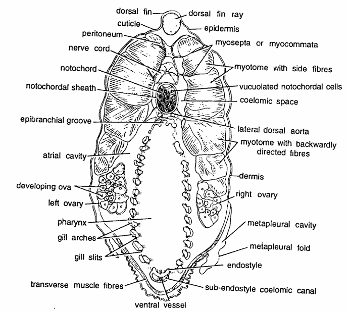
Amphioxus Slides : T.S. Passing Through Ovaries
Comments
Under low magnification : (10 X eye-piece; 4 X objective).
- T.S. passing through above region shows body-wall layers, nerve cord, notochord, pharynx, midgut (liver) and ovaries.
- Body wall is composed of cuticle, epidermis, dermis and muscle layer. Cuticle, epidermis and dermis are very thin and indistinguishable.
- The musculature consisting of longitudinal fibers is well developed.
- First four segmental myotomes are thick and separated by myosepta.
- Last two myotomes are comparatively thinner.
- Muscle fibres in first three myotomes are directed sideways and upwards while muscle fibres in last three myotomes are backwardly directed.
- Dorsal tin ray is present just beneath the mid-dorsal epidermis.
- Below fin ray is nerve cord containing neurocoel.
- Notochord below nerve cord with chordal sheath enclosing vacuolated notochordal cells filled with homogeneous fluid.
- Ventral part of section contains two ovaries.
- Ovaries, enclosed in coelomic sac, contain several ova and are found from 25-51 segments. Pharynx, surrounded by atrial cavity, contains gill slits, epipharyngeal groove dorsally and endostyle ventrally.
- Two metapleural folds are seen ventro-Iaterally. In some sections midgut diverticulum or liver is also seen.
- Other structures seen are ventral vessel sub-endostyler coelomic caudal and coelomic space.
Identification: Since the section contains ova and all above characters, hence it is T.S. of female through ovaries of Amphioxus.
Amphioxus Slides : T.S. Passing Through Testes
Comments
- T.S. passing through testes shows body wall layers, nerve cord, notochord, large pharynx, etc.
- Body wall is composed of thin cuticle, thin epidermis and dermis and thick cut longitudinal segmental myotomes separated by myosepta.
- First four myotomes are quite thick and last two comparatively thinner.
- Muscle fibres in first three myotomes are slightly upwardly directed while in posterior three backwardly directed.
- Dorsal tin ray just below epidermis. Nerve cord containing neurocoel is present below the fin ray.
- Below the nerve cord is present notochord composed of chordal sheath enclosing vacuolated notochordal cells filled with homogeneous fluid.
- Pharynx is a large cavity and contains dorsal epipharyngeal groove, ventral endostyle and on sides several gill clefts and gill arches.
- Testes are found on both sides having several dot-shaped cut spermatozoa.
- Metapleural folds and metapleural canals are seen ventro laterally.
- Other structures seen are gill arches, peritoneum, transverse muscle fibres, sub-endostyler coelomic canal, coelomic spaces, dorsal aorta, septum and ocellus.
Identification: Since the section contains dot-shaped cut spermatozoa, and all above characters, hence it is T.S. of male through testes of Amphioxus.
T. S. Passing Through Mid-gut or Intestine
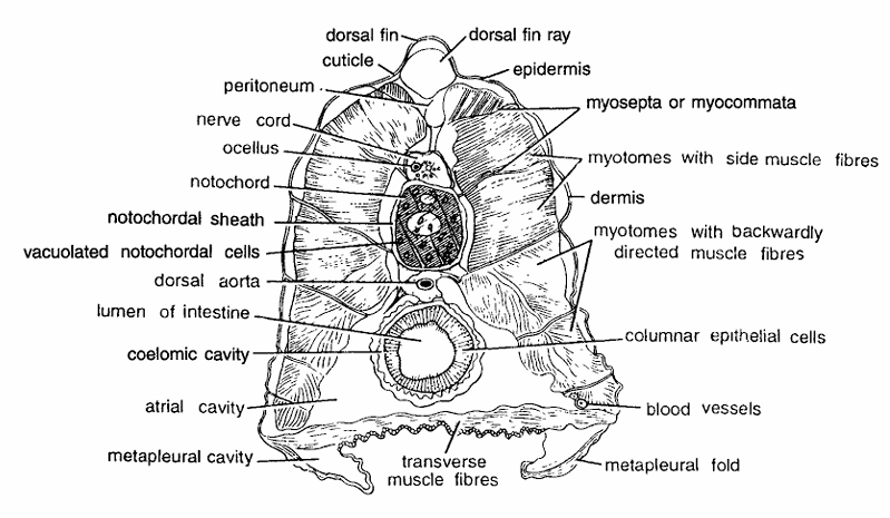
Comments
- Intestine is found in posterior region. T.S. through intestine shows usual body wall, layers, nerve cord, notochord, intestine and metapleural folds.
- Body wall is composed of thin cuticle, thin epidermis, dermis and muscle layer.
- Muscle layer consists of alternating thick segmental myotomes separated by myocommata.
- Four myotomes forming anterior end are very thick while posterior last three myotomes are thinner.
- Muscle fibres in first three myotomes are directed slightly upwards while last three have backwardly directed muscle fibres.
- Dorsal fin ray is found below epidermis. Glandular nerve cord is found below dorsal fin ray. It encloses central canal or neurocoel.
- Notochord found below nerve cord is with vacuolated chordal cells. There is a single dorsal aorta below notochord.
- Ventral half of the section contains mid-gut, coelomic cavity and atrial cavity. Below coelom is well developed atrial cavity.
- Below atrial cavity transverse muscles and metapleural folds are seen.
- Mid-gut or intestine is found in the centre, composed of large endodermal columnar ciliated epithelial cells.
- Other structures seen in the section one ocellus, peritoneum, metapleural cavity, transverse muscle fibres, metapleural folds, blood vessels.
Identification : Since the section contains cut intestine, renal papillae and all above characters, hence it is T.S. of Amphioxus through intestine.
T.S. Passing Through Atriopore
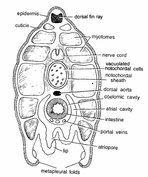
Comments
- Animal narrows towards the posterior region. Hence T.S. passing through atriopore shows smaller sectIOn. In the section body wall layers, nerve cord, notochord, and atriopore are seen.
- Body wall comprises of thin cuticle, single-layered thin epidermis, thin dermis and segmental muscles or myotomes which are comparatively thinner and separated by myocommata.
- Dorsal fin ray is present below the epidermis.
- Nerve cord is found below 2 or 3 myotomes in the middle. It contains neurocoel.
- Notochord with notochordal sheath and vacuolated chordal cells is present below the nerve cord.
- Just below notochord is single dorsal aorta. Intestine shows smaller diameter and is composed of endodermal cells.
- It is surrounded by coelomic and atrial cavities.
- Few cut portal veins are also seen. Atrial cavity surrounds coelom and opens ventrally by a distinct atriopore, situated in front of the ventral fin.
- The two metapleural folds are distinctly seen.
Identification : Since this section contains atriopore and all above characters, hence it is T.S. of Amphioxus through atriopore.
T.S. Passing Through Anal Region
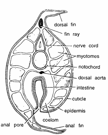
Comments
- As the body narrows posteriorly the T.S. passing through anal region shows smaller diameter and it tapers at both the ends.
- Body wall is composed of thin cuticle, epidermis, dermis and myotomes alternating with myosepta.
- Dorsal fin ray is present below the pointed epidermis dorsally.
- Nerve cord is found below dorsal fin ray and first myotome.
- It has central canal. Notochord is found beneath the nerve cord.
- It has vacuolated chordal cells. Dorsal aorta is present beneath the notochord.
- Coelomic cavity enclosing intestine. Intestine opens to the exterior by the anus. Ventral fin is pointed in section.
Identification : Since the section contains anus and all above characters, hence it is T.S. of Amphioxus through anal region.
T.S. Passing Through Caudal Region
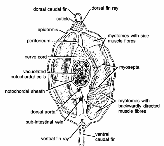
Comments
- Section through caudal region is somewhat smaller in size and without any opening.
- Body wall is composed of thin cuticle, single-layered epidermis, dermis and myotomes alternating with myocommata.
- Three upper myotomes have side muscle fibres while 2 posterior ores have backwardly directed fibers.
- Myotomes are separated by myosepta.
- Dorsal fin ray found at the base of dorsal fin below epidermis.
- Nerve cord with neurocoel lies below dorsal fin ray.
- Notochord with vacuolated chordal cells is found below the nerve cord.
- Caudal artery and vein appear below notochord.
- Alimentary canal, atrial cavity, coelom and metapleural folds are absent in this section.
- Caudal fin with fin ray is present posteriorly.
Identification : Since there is no opening, it is T.S. passing through the caudal region of Amphioxus.
Discover more from Zoologyverse
Subscribe to get the latest posts sent to your email.
Pingback: BRANCHIOSTOMA LANCEOLATUM | Zoologyverse