Placentation in Mammals
What is Placenta
Modes of embryonic nutrition differ in different mammals. Prototheria or monotremes (Tachyglossus, Ornithorhynchus) are oviparous like most reptiles and birds. They produce large, heavily yolked and shelled eggs. The vitelline vessels developed in the wall of yolk sac carry yolky nutrients to the developing embryo. There is no uterine gestation and no formation of placenta. Newly hatched young are exceedingly immature and complete their growth in a temporarily developed abdominal pouch of mother during which they are fed upon milk.
All other mammals (Metatheria and Eutheria) are viviparous. The development of their young is intra-uterine, that is, inside the uterus of mother. But their minute eggs contain so little yolk that they could never develop beyond the very early stages unless additional nourishment is somehow provided by the mother. This is done by the formation of a characteristic organ called placenta by which the embryos establish close contact with the utrine wall of mother. The term placenta may be defined as the structure by which the developing embryo or foetus of viviparous mammals obtains its nourishment from the maternal uterine blood. It is formed by the interlocking of both foetal as well as maternal tissues. The part derived from foetus is called foetal placenta, while that derived from uterine wall is called maternal placenta. The term placentation may be defined as an intimate relation between a portion of maternal uterine wall and a part or whole of the chorionic membrane or trophoblast of embryo for the purpose of nutrition, respiration and excretion. Placentation involves a series of events following implantation of embryo and leading to development of placenta. Placenta is not exclusively found in mammals but also present in other animal groups like— Onychophora (Peripatus), Ascidians (Salpa), Elasmobranchs (Mustelus) and some lizards also. But the tissues which help in formation of placenta are different from those of mammals.
Implantation of Embryo
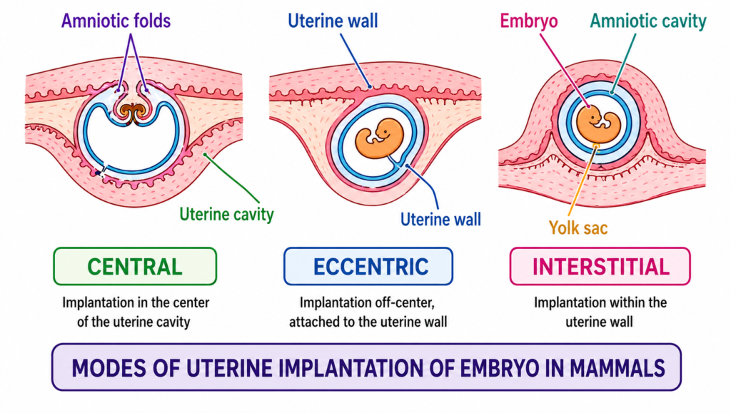
First step towards formation of placenta is the attachment of developing embryo to the wall of uterus, called implantation. This is effected by small finger-like processes, the villi, which grow out from a particular area or areas of trophoblast of embryo and penetrate into corresponding crypts in the maternal uterine wall. Blood vessels, lymphatic vessels and glands of uterine wall enlarge and their secretions in the lumen of uterus provide nourishment to the embryo. Implantation generally occurs in one of the following three manners
- Central implantation :- In rabbit, ungulates, carnivores and lower primates, embryo is attached to the surface of uterine lining and projects freely into uterine cavity. This is also known as superficial implantation.
- Concentric implantation :- In mouse, rat, squirrel, beaver, etc. the mucous lining of uterus gives out folds to cover the embryo which is embedded in a groove or pocket of main uterine cavity.
- Interstitial implantation :- In man, apes, pig and certain rodents such as guinea pig and hedgehog, the blastocyst or embryo actually burrows into uterine tissue to become completely surrounded by it.
As far as mechanism of implantation is concerned, when the blastocyst comes in the direct contact of the uterine epithelium, in the area of attachment, begins to break down by the action of some digestive enzymes secreted by trophoblast. The erosion of uterine epithelium creates gap through which invading trophoblast advances and comes into contact with the connective tissue layer of uterus. The secretion of progesterone by the corpus luteum makes the endometrium more receptive for implantation.
Classification of Placenta
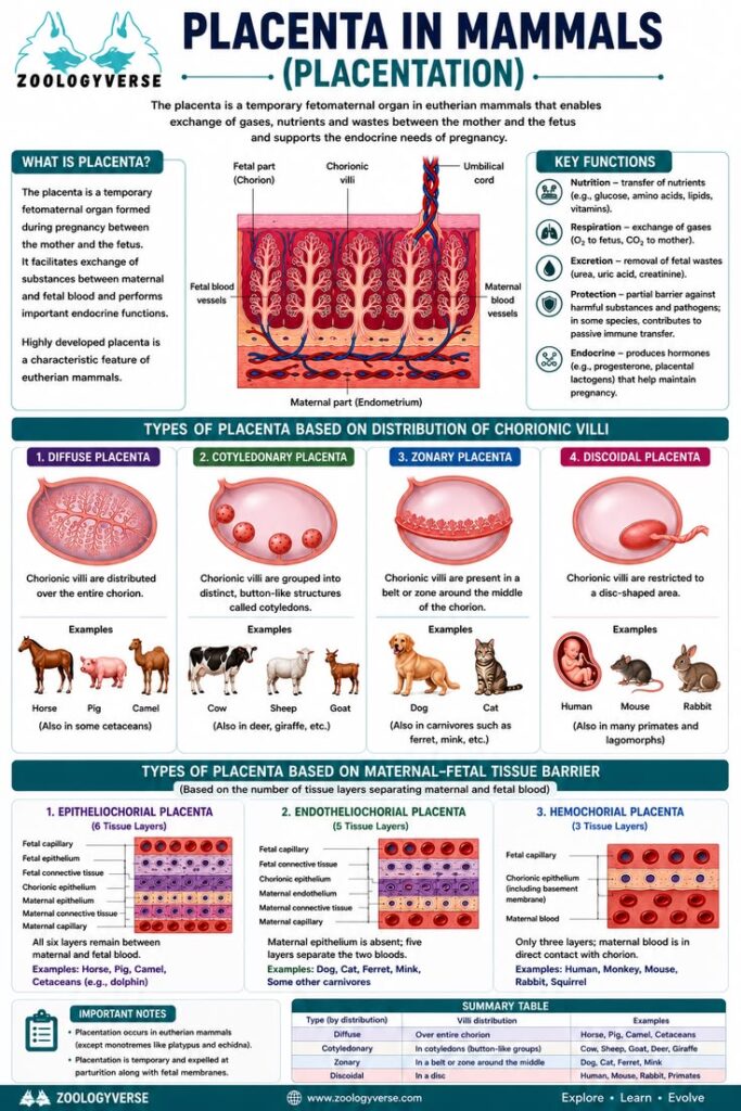
Mammals show many variations in the mode of origin and details of shape and structure of placenta, which are classified accordingly. The three main factors involved are : (i) Nature of extra-embryonic membranes involved, (ii) distribution of villi and shape of placenta, and (iii) degree of intimacy between foetal and maternal tissues or histology.
(I)Types according to extra-embryonic membranes involved or mode of origin
Depending on the foetal membranes forming placenta, three kinds are recognized : yolk sac, allantoic and chorionic.
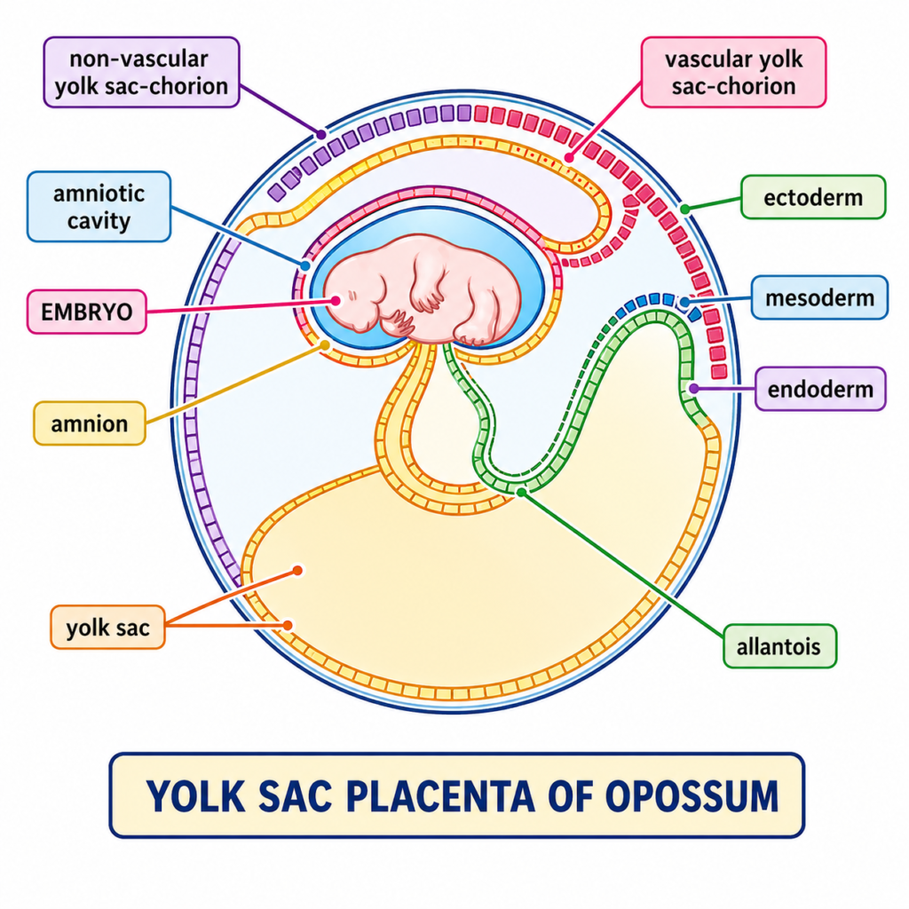
1.Yolk sac placenta :- In Metatheria or marsupials, such as kangaroo (Macropus) and opossum (Didelphys), placenta is derived from yolk sac and chorion. Yolk sac developed from the lower part of blastocyst is very large and nearly encloses the entire embryo and its amnion. Wall of yolk sac lies in direct contact with chorion (trophoblast) which sends out finger-like villi into uterine wall. Yolk sac wall also develops vitelline blood vessels for transporting secretions. Uterine milk absorbed from uterus to the developing embryo. Allantois remains poorly developed and never comes in contact with chorion.
In Metatheria, yolk sac placenta is only weakly developed so that embryonic nutrition and growth remain limited and the young is born very small and immature. To compensate the deficiency of intra-uterine development, it is transferred to the abdominal pouch or marsupium and fed on milk until fully formed.
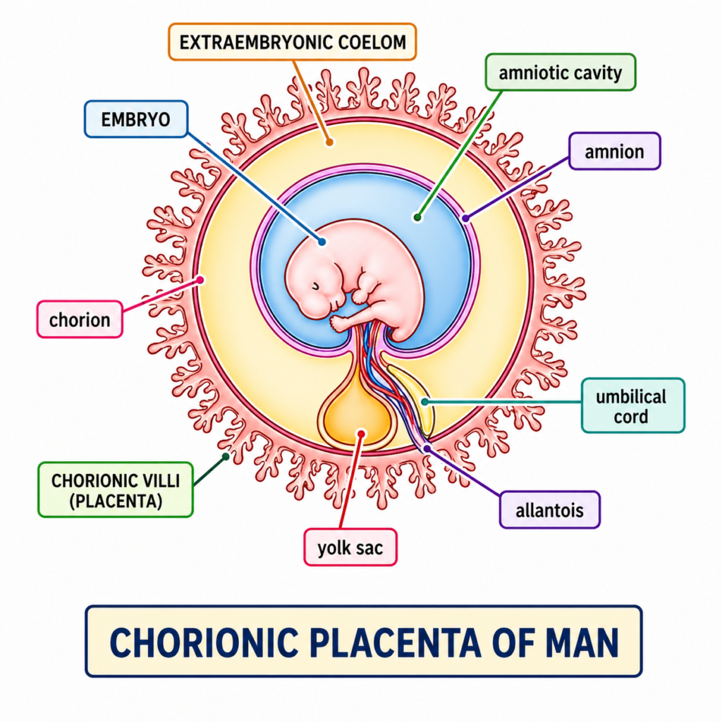
In higher mammals (Eutheria), a yolk sac placenta is usually not found. But, it may be large and temporarily develop in early stages in some mammals such as hedgehogs and rabbits. Or, it may be small ending in a small tube in the umbilical cord, as in man.
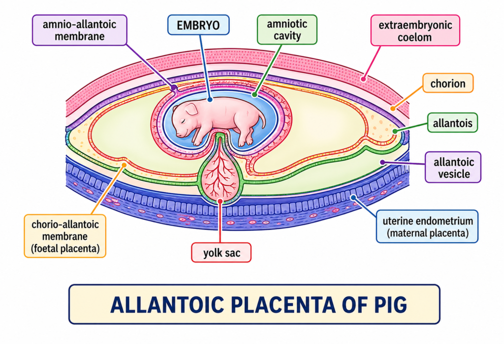
2. Allantoic placenta :- In the majority of Eutheria, the chief organ of embryonic nutrition is the allantoic placenta consisting of allantois and chorion. Allantois is a sac-like outgrowth from the hindgut of embryo. It is lined internally by endoderm and externally by mesoderm. As allantois grows and spreads in the extra-embryonic cavity, its mesoderm fuses with that of chorion over a somewhat restricted region. The layer formed by fusion of allantois and chorion is termed allanto-chorion. It becomes richly vascular and thrown into small, finger-like processes, the villi. The uterine wall forms corresponding depressions, called crypts, which are penetrated by foetal villi forming allantoic placenta. Materials absorbed from maternal blood through allantoic placenta are carried to the foetus by allantoic blood vessels. Outside Eutheria, a primitive allantoic placenta occurs only in Perameles (bandicoot) which is a metatherian. But it also has an efficient yolk sac placenta. In this case yolk sac and allantois are large, well developed but it is allantois that supplies blood vessels to chorion. The trophoblast of the chorion, at places of contact with uterine wall disappears. The uterine wall is syncytial and highly vascularised. Physiological exchange takes place between the foetal blood and maternal blood.
3. Chorionic placenta :- It occurs in man and apes and is formed only by chorion. Allantois remains small, burrows into body stalk (umbilicai cord) and does not reach chorion. However, its mesoderm and blood vessels grow up to chorion whose villi enter the uterine crypts forming chorionic placenta.
[II] Types according to shape and distribution of villi
Depending on the shape of placenta, manner or distribution of villi, degree of connection between foetal and maternal tissues and behaviour of placenta at the time of birth, the following types and subtypes of allantoic placenta can be recognized :-
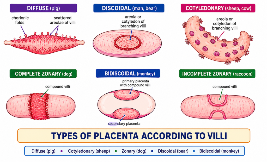
- Non-deciduous placenta. In most mammals villi are simple, unbranched and merely apposed without intimate contact between foetus and uterine wall. At the time of birth or parturition, villi are easily withdrawn from maternal crypts without causing any tissue damage. Thus no part of uterine tissue comes out and no bleeding occurs. Non-deciduous or non-deciduate placenta has following subtypes according to the manner of distribution of villi.
- Deciduous placenta :- Villi are complicated, branched and intimately connected. At birth, a variable amount of maternal tissue is pulled out with the shedding of blood. Deciduous or deciduate placenta is also differentiated in the following subtypes
- Zonary :- Villi form an incomplete (e.g. racoon) complete girdle encircling blastocyst. Ex. cat, dog, seal, elephant.
- Discoidal :- Villi are restricted to a circular disc or plate on the dorsal surface of blastocyst. Ex. insectivores, bats, rodents (rat, mouse), rabbit, bear.
- Metadiscoidal :- Villi are at first scattered but later become restricted to one or two discs. It is monodiscoidal in man and bidiscoidal in monkeys and apes
- Contra-deciduous :- Foetal villi and uterine crypts are so intimately connected that even most of foetal placenta is left behind at birth to be broken and absorbed by maternal leucocytes. Ex. Bandicoot (Perameles), mole (Talpa).
[Ill] Histological types
Foetal and maternal bloods in placenta do not mix up with each other. To start with, the two blood streams are separated from each other by atleast the following 6 tissue barriers or membranes :
- Endothelium of maternal blood vessels
- Uterine connective tissue
- Uterine epithelium
- Chorionic epithelium or trophoblastic ectoderm
- Chorionic + allantoic mesoderm
- Endothelium of foetal blood vessels
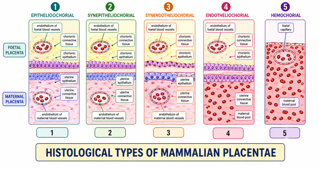
Exchange of substances in solution between two blood streams occurs by diffusion through these tissues. To increase effectiveness or efficiency of placenta, it is necessary to reduce the number of tissues between foetal and maternal blood streams. Nature of reduction or erosion of tissues varies greatly in different mammals. Grossor recognizes the following 5 histological types or grades of reduction
- Epithelio-chorial. It is the simplest type with all the six tissue barriers present. It is supposed to be most primitive from which other types have been derived. Ex. Pigs, lemurs.
- Syndesmo-chorial. Only uterine epithelium is eroded so that chorionic epithelium (or trophoblastic ectoderm) comes in contact with uterine connective tissue. Ex. Cattle, sheep.
- Endothelio-chorial. Uterine epithelium as well as uterine connective tissue breakdown so that chorionic epithelium comes in contact with the endothelium of maternal blood vessels. Ex. Carnivore (cat, dog), tree shrew, mole.
- Haemo-chorial. Endothelium of maternal blood vessels also disappears. Thus chorionic epithelium and blood vessels of foetal villi are directly bathed in maternal blood flowing in sinuses. Ex. Man, apes, monkeys, some insectivores and some rodents.
- Haemo-endothelial. Finally, the chorionic epithelium and mesoderm also disappear allowing the foetal capillaries to come in direct contact with maternal blood. Ex. Rat, guinea pig, rabbit.
On the basis of electron microscopic studies of the histology of placenta, placenta can be classified into Epithelio-chorial three categories placenta, only viz., Endotheliochorial placenta and Haemochorial placenta. Goel (1984) further classified haemochorial type of placenta into three subtypes — haemomonochorial type, haemodichorial type and haemotrichorial type, based on the number of layers of trophoblast involved in covering the foetal endothelium.
Physiology and Functions of Placenta
Placentation is the mechanism by which the foetal and maternal blood circulations are brought very close together to provide for the respiration, excretion and nutrition of the foetus. However, there is no mixture or fusion of these two blood streams. Foetal blood does not circulate in mother and vice versa. Exchange of substances occurs from one circulation to the other by diffusion in which the intervening tissue barriers serve as an ultrafilter. Only selected substances can pass through placenta. All food materials and oxygen of maternal blood diffuse into foetal capillaries, while excretory wastes and carbon dioxide produced by foetus diffuse in the opposite direction. In addition, placenta stores materials such as fat, glycogen and iron for the use of embryo while it still has no liver. Placenta also participates in the metabolism of proteins and serves as an endocrine gland. In rabbit, placenta secretes a protein hormone, relaxin, which causes relaxation of pelvic ligaments and pubic symphysis to facilitate birth of the young. In human and other placental mammals it secretes placental lactogen, chorionic gonadotropin, estradiol, progesterone, etc. Besides these, significant immunological function has also been reported by the trophoblast (Dutta, 1987).
Placenta and Diseases
Viral or bacterial infection of placenta is known as placentitis. If the mother suffers from certain diseases like syphilis, small pox, chicken pox, measles, etc., their viruses or pathogenic organisms may pass through placenta to infect the foetus with these diseases. Drugs such as thalidomide taken as a sedative by mother during early pregnancy, may effect serious deficiency in limbs, heart or digestive tract of foetus. Normally maternal blood does not come in direct contact with foetal blood. However, accidental breaking of placental capillaries may result in some red blood cells of foetus entering maternal circulation. If per chance mother is Rh negative and foetus is Rh positive, the positive red cells of foetus will induce production of Rh antibody in maternal blood plasma, which can easily diffuse through into foetal circulation damaging its red cells, and resulting in jaundice or even death of foetus.
On the other hand, if mother has acquired immunity against certain diseases like diphtheria, small pox, measles, etc., the antibodies developed in maternal blood are passed to foetus which also acquires immunity against such diseases.
Discover more from Zoologyverse
Subscribe to get the latest posts sent to your email.